. 2003 May;23(9):3103-15.
doi: 10.1128/MCB.23.9.3103-3115.2003.

Transmodulation between phospholipase D and c-Src enhances cell proliferation

Affiliations

Transmodulation between phospholipase D and c-Src enhances cell proliferation

Bong-Hyun Ahn et al. Mol Cell Biol. 2003 May.

Abstract

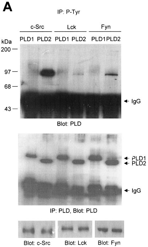
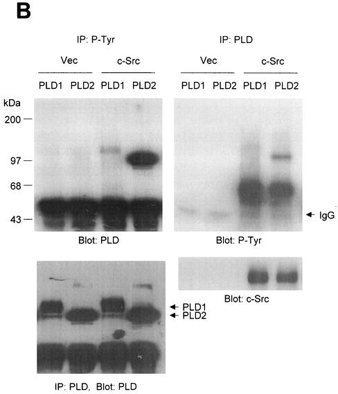
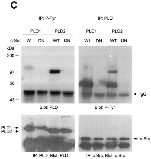
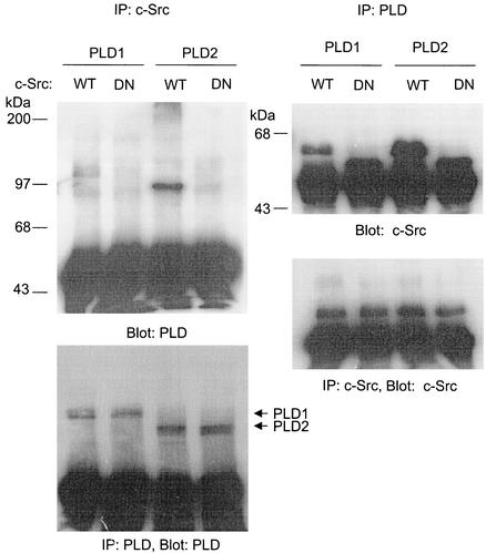
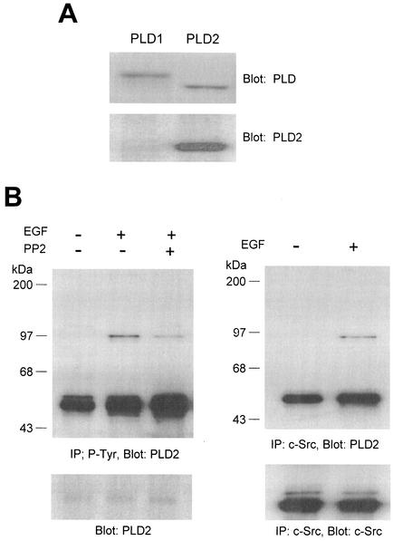
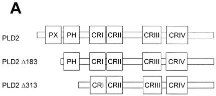
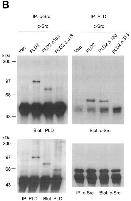
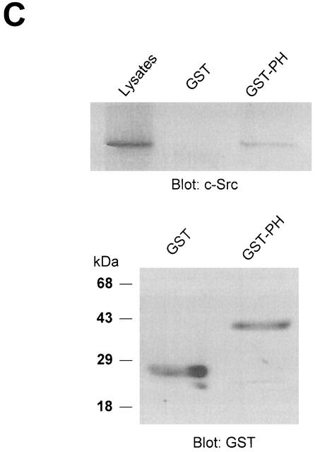
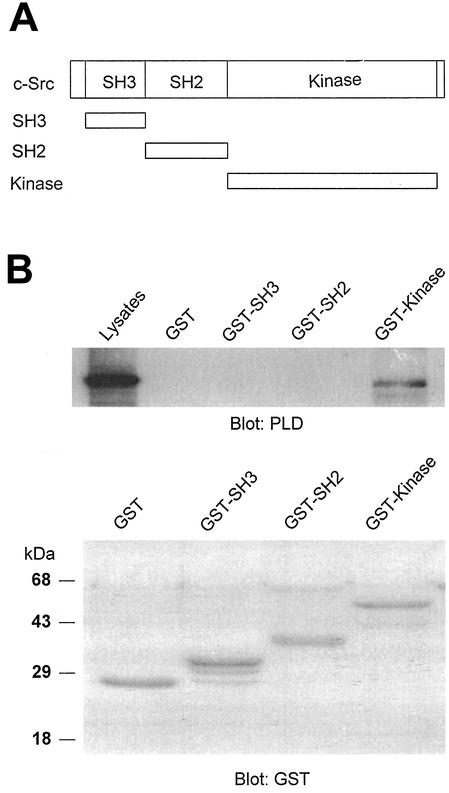
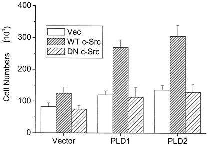
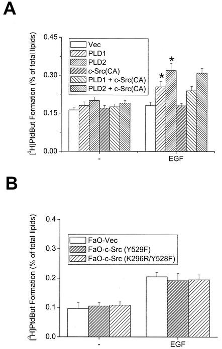
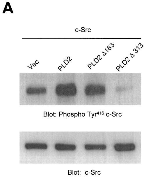
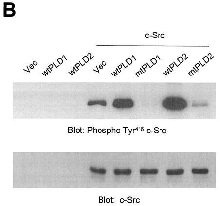
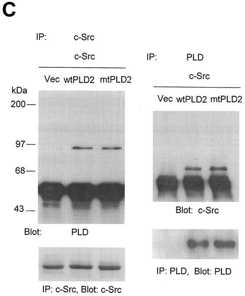
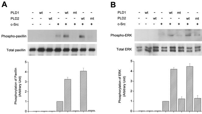
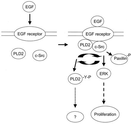
Phospholipase D (PLD) has been implicated in the signal transduction pathways initiated by several mitogenic protein tyrosine kinases. We demonstrate for the first time that most notably PLD2 and to a lesser extent the PLD1 isoform are tyrosine phosphorylated by c-Src tyrosine kinase via direct association. Moreover, epidermal growth factor induced tyrosine phosphorylation of PLD2 and its interaction with c-Src in A431 cells. Interaction between these proteins is via the pleckstrin homology domain of PLD2 and the catalytic domain of c-Src. Coexpression of PLD1 or PLD2 with c-Src synergistically enhances cellular proliferation compared with expression of either molecule. While PLD activity as a lipid-hydrolyzing enzyme is not affected by c-Src, wild-type PLDs but not catalytically inactive PLD mutants significantly increase c-Src kinase activity, up-regulating c-Src-mediated paxillin phosphorylation and extracellular signal-regulated kinase activity. These results demonstrate the critical role of PLD catalytic activity in the stimulation of Src signaling. In conclusion, we provide the first evidence that c-Src acts as a kinase of PLD and PLD acts as an activator of c-Src. This transmodulation between c-Src and PLD may contribute to the promotion of cellular proliferation via amplification of mitogenic signaling pathways.


Figures

FIG. 1.
FIG. 1.
PLD2 and, to a lesser extent, PLD1 are tyrosine phosphorylated by c-Src tyrosine kinase. (A) COS-7 cells were transiently cotransfected for 40 h with various combinations of plasmids encoding PLD1, PLD2, c-Src, Lck, and Fyn. Cell lysates were immunoprecipitated with anti-P-Tyr antibody, and precipitated proteins were subjected to immunoblot analysis with an anti-PLD antibody. Western blot analysis using PLDs and c-Src family tyrosine kinase demonstrated that the respective proteins were expressed to equal extents. (B) COS-7 cells were transiently transfected with the indicated expression plasmids, and cell lysates were subjected to immunoprecipitation with anti-P-Tyr antibody or anti-PLD antibodies. Immunoprecipitates were separated by SDS-PAGE and immunoblotted with anti-PLD or anti-P-Tyr antibody. Expression of PLD and c-Src was determined by using anti-PLD or anti-c-Src antibodies. (C) COS-7 cells were transiently transfected with various combinations of plasmids encoding PLD1, PLD2, wild-type (WT) c-Src, and dominant negative (DN) c-Src. Cell lysates were immunoprecipitated by using anti-P-Tyr antibody or anti-PLD antibody, and immune complexes were analyzed by immunoblotting. Immunoreactive proteins were visualized by use of horseradish peroxidase-coupled secondary antibody and chemiluminescence. Data are representative of three experiments.
FIG. 1.
FIG. 1.
PLD2 and, to a lesser extent, PLD1 are tyrosine phosphorylated by c-Src tyrosine kinase. (A) COS-7 cells were transiently cotransfected for 40 h with various combinations of plasmids encoding PLD1, PLD2, c-Src, Lck, and Fyn. Cell lysates were immunoprecipitated with anti-P-Tyr antibody, and precipitated proteins were subjected to immunoblot analysis with an anti-PLD antibody. Western blot analysis using PLDs and c-Src family tyrosine kinase demonstrated that the respective proteins were expressed to equal extents. (B) COS-7 cells were transiently transfected with the indicated expression plasmids, and cell lysates were subjected to immunoprecipitation with anti-P-Tyr antibody or anti-PLD antibodies. Immunoprecipitates were separated by SDS-PAGE and immunoblotted with anti-PLD or anti-P-Tyr antibody. Expression of PLD and c-Src was determined by using anti-PLD or anti-c-Src antibodies. (C) COS-7 cells were transiently transfected with various combinations of plasmids encoding PLD1, PLD2, wild-type (WT) c-Src, and dominant negative (DN) c-Src. Cell lysates were immunoprecipitated by using anti-P-Tyr antibody or anti-PLD antibody, and immune complexes were analyzed by immunoblotting. Immunoreactive proteins were visualized by use of horseradish peroxidase-coupled secondary antibody and chemiluminescence. Data are representative of three experiments.
FIG. 2.
FIG. 2.
PLD isozymes, most notably PLD2, bind to wild-type c-Src but not to the kinase-inactive c-Src mutant. COS-7 cells were transiently transfected for 40 h with various combinations of plasmids encoding PLD1, PLD2, wild-type (WT) c-Src, and dominant negative (DN) c-Src. Cell lysates were immunoprecipitated with anti-PLD or anti-c-Src antibodies, and immune complexes were subjected to immunoblotting analysis using anti-c-Src or anti-PLD antibodies, respectively. Expressions of PLDs and c-Src were determined by immunoprecipitation and immunoblotting. Data are representative of three experiments.
FIG. 3.
FIG. 3.
EGF induces tyrosine phosphorylation of PLD2 and its association with c-Src in A431 cells. (A) Mouse monoclonal antibody specific for PLD2 was generated as described in Materials and Methods. To analyze its specificity, PLD1 or PLD2 was transfected into COS-7 cells and the lysates were immunoblotted with anti-PLD antibody which recognizes both PLD1 and PLD2 or anti-PLD2 antibody. (B) Quiescent A431 cells were pretreated with or without a selective inhibitor of Src family tyrosine kinase, PP2 (2 μM), for 30 min and stimulated with 200 ng of EGF/ml for 20 min. Cell lysates were prepared, and 1 mg of the lysates were immunoprecipitated with anti-P-Tyr antibody or anti-c-Src antibody. Immunoprecipitated proteins were separated on SDS-PAGE gel and probed with mouse monoclonal antibody specific for PLD2. The result shown is representative of three experiments.
FIG. 4.
FIG. 4.
The N-terminal PH domain of PLD2 is required for interaction with c-Src. (A) Schematic representation of PLD2. The boxes indicate highly conserved sequences of PLD; their possible functions have been proposed or demonstrated in reference . CR, conserved region. (B) COS-7 cells were transiently cotransfected with plasmids encoding the empty vector (Vec) and c-Src, or PLD2 and c-Src, or N-terminal 183-amino-acid-truncated PLD2 Δ183(184-933) and c-Src, or N-terminal 313-amino-acid-truncated PLD2 Δ313(314-933) and c-Src. Cell lysates were subjected to immunoprecipitation by using anti-PLD or anti-c-Src antibodies, and the amount of coimmunoprecipitated c-Src or PLD was determined by immunoblotting with anti-c-Src or anti-PLD antibodies. Expression of PLD2 and c-Src was determined by using anti-PLD or anti-c-Src antibodies. (C) For the GST pull-down assay, GST-PH fusion protein was used as described in reference . The lysates transfected with c-Src were incubated with 2 μg of GST or GST-PH fragment and immunoblotted with antibody to c-Src. The amount of the GST fusion proteins was visualized by immunoblotting with an anti-GST antibody. Data are representative of three experiments.
FIG. 4.
FIG. 4.
The N-terminal PH domain of PLD2 is required for interaction with c-Src. (A) Schematic representation of PLD2. The boxes indicate highly conserved sequences of PLD; their possible functions have been proposed or demonstrated in reference . CR, conserved region. (B) COS-7 cells were transiently cotransfected with plasmids encoding the empty vector (Vec) and c-Src, or PLD2 and c-Src, or N-terminal 183-amino-acid-truncated PLD2 Δ183(184-933) and c-Src, or N-terminal 313-amino-acid-truncated PLD2 Δ313(314-933) and c-Src. Cell lysates were subjected to immunoprecipitation by using anti-PLD or anti-c-Src antibodies, and the amount of coimmunoprecipitated c-Src or PLD was determined by immunoblotting with anti-c-Src or anti-PLD antibodies. Expression of PLD2 and c-Src was determined by using anti-PLD or anti-c-Src antibodies. (C) For the GST pull-down assay, GST-PH fusion protein was used as described in reference . The lysates transfected with c-Src were incubated with 2 μg of GST or GST-PH fragment and immunoblotted with antibody to c-Src. The amount of the GST fusion proteins was visualized by immunoblotting with an anti-GST antibody. Data are representative of three experiments.
FIG. 5.
FIG. 5.
The catalytic domain of c-Src interacts with PLD2. (A) Schematic representation of GST-fusion protein constructs. (B) c-Src was fragmented into SH3 (88-137), SH2 (144-245), and kinase (265-523) domains. The fragments were cloned as GST fusion proteins, expressed in Escherichia coli, and purified by use of glutathione-conjugated Sepharose beads. An equal amount (2 μg) of GST or GST-Src fragment was incubated with the lysates of COS-7 cells transfected with PLD2 and c-Src. The pull-down proteins were subjected to immunoblot analysis with an antibody against PLD (upper panel). The amount of the GST fusion proteins was visualized by immunoblotting with an anti-GST antibody (lower panel). Data are representative of three experiments.
FIG. 6.
FIG. 6.
Modulation between PLD2 and c-Src significantly enhances cellular proliferation. Mouse fibroblasts overexpressing PLD isozymes were plated at a density of 5 × 10⁴ cells in 12-well plates and transfected with empty vector (Vec), wild-type (WT) c-Src, or dominant negative (DN) c-Src mutant. After 40 h of incubation, viable cells were counted by a trypan blue exclusion method. Results show means ± standard deviations of three independent experiments.
FIG. 7.
FIG. 7.
c-Src does not affect EGF-induced PLD activation in COS-7 and FaO cells. COS-7 cells transiently transfected with various constructs (A) and FaO cells expressing constitutive active c-Src (Y529F) or dominant inactive c-Src mutant (K296R/Y528F) (B) were labeled with [3H]myristic acid for 18 h. The cells were untreated or treated with 200 ng of EGF/ml for 30 min in the presence of 0.3% 1-butanol. Radioactivity incorporated into PtdBut was measured as described in Materials and Methods. *, P < 0.05 compared to cells transfected with vector and treated with EGF. Results show means ± standard deviations of three independent experiments.
FIG. 8.
FIG. 8.
Overexpression of PLD stimulates c-Src kinase activity. (A) COS-7 cells were transiently cotransfected with various combinations of plasmids encoding empty vector (Vec) and c-Src, or PLD2 and c-Src, or N-terminal 183-amino-acid-truncated PLD2 Δ183(184-933) and c-Src, or N-terminal 313-amino-acid-truncated PLD2 Δ313(314-933) and c-Src. Cell lysates were subjected to immunoblot analysis using a phospho Tyr416 c-Src antibody. Expression of c-Src was determined by using an anti-c-Src antibody. (B) COS-7 cells were transiently transfected for 40 h with various combinations of plasmids encoding an empty vector, PLD1, PLD2, c-Src, and c-Src plus either wild-type (wt) or catalytically inactive mutant (mt) PLD1 or PLD2. Data are representative of three experiments. (C) COS-7 cells were transiently cotransfected with a catalytically inactive PLD2 mutant and wild-type c-Src, and cell lysates were subjected to immunoprecipitation with anti-c-Src antibody or anti-PLD antibodies. Immunoprecipitates were separated by SDS-PAGE and immunoblotted with anti-PLD or anti-c-Src antibody. Expression of PLD and c-Src was determined by using anti-PLD or anti-c-Src antibodies. Data are representative of three experiments.
FIG. 8.
FIG. 8.
Overexpression of PLD stimulates c-Src kinase activity. (A) COS-7 cells were transiently cotransfected with various combinations of plasmids encoding empty vector (Vec) and c-Src, or PLD2 and c-Src, or N-terminal 183-amino-acid-truncated PLD2 Δ183(184-933) and c-Src, or N-terminal 313-amino-acid-truncated PLD2 Δ313(314-933) and c-Src. Cell lysates were subjected to immunoblot analysis using a phospho Tyr416 c-Src antibody. Expression of c-Src was determined by using an anti-c-Src antibody. (B) COS-7 cells were transiently transfected for 40 h with various combinations of plasmids encoding an empty vector, PLD1, PLD2, c-Src, and c-Src plus either wild-type (wt) or catalytically inactive mutant (mt) PLD1 or PLD2. Data are representative of three experiments. (C) COS-7 cells were transiently cotransfected with a catalytically inactive PLD2 mutant and wild-type c-Src, and cell lysates were subjected to immunoprecipitation with anti-c-Src antibody or anti-PLD antibodies. Immunoprecipitates were separated by SDS-PAGE and immunoblotted with anti-PLD or anti-c-Src antibody. Expression of PLD and c-Src was determined by using anti-PLD or anti-c-Src antibodies. Data are representative of three experiments.
FIG. 9.
FIG. 9.
PLD enhances not only c-Src-induced tyrosine phosphorylation of paxillin but also c-Src-mediated ERK activation. COS-7 cells were transiently cotransfected for 40 h with various combinations of plasmids encoding an empty vector, PLD1, PLD2, c-Src, and c-Src plus either wild-type (wt) or catalytically inactive mutant (mt) PLD1 or PLD2. Cell lysates were immunoblotted with an antibody directed against tyrosine-phosphorylated paxillin (A) or an anti-phospho-ERK antibody (B). Expression of total paxillin and ERK was visualized by immunoblotting with antipaxillin antibody or anti-ERK antibody. Data are representative of three experiments. The intensity of phosphorylated paxillin or phospho-ERK immunoreactive bands quantified by densitometry of immunoblot was expressed as relative intensity of the bands. Results show means ± standard deviations of three independent experiments.
FIG. 10.
FIG. 10.
Proposed model of PLD-stimulated c-Src downstream signaling. EGF binds to its receptor, thereby stimulating both PLD and c-Src activation. It is likely that the resulting PLD activity is involved in the stimulation of Src-mediated signaling pathways such as ERK and then in the stimulation of cell proliferation. EGF induces tyrosine phosphorylation of PLD2 via c-Src and its interaction with c-Src. At present, it is not clear, however, how tyrosine phosphorylation of PLD is involved in PLD-mediated signaling mechanism.

Similar articles

Cited by

References

    1. Ahn, B. H., H. Rhim, S. Y. Kim, Y. M. Sung, M. Y. Lee, J. Y. Choi, B. Wolozin, J. S. Chang, Y. H. Lee, T. K. Kwon, K. C. Chung, S. H. Yoon, S. J. Hahn, M. S. Kim, Y. H. Jo, and D. S. Min. 2002. α-Synuclein interacts with phospholipase D isozymes and inhibits pervanadate-induced phospholipase D activation in human embryonic kidney-293 cells. J. Biol. Chem. 277:12334-12442. - PubMed
    1. Ben-Av, P., and M. Liscovitch. 1989. Phospholipase D activation by the mitogens platelet-derived growth factor and 12-O-tetradecanoylphorbol 13-acetate in NIH-3T3 cells. FEBS Lett. 259:64-66. - PubMed
    1. Bjorge, J. D., A. Jakymiw, and D. J. Fujita. 2000. Selected glimpses into the activation and function of Src kinase. Oncogene 19:5620-5635. - PubMed
    1. Bradford, M. M. 1976. A rapid and sensitive method for the quantitation of microgram quantities of protein utilizing the principle of protein-dye binding. Anal. Biochem. 72:248-254. - PubMed
    1. Brown, M. T., and J. A. Cooper. 1996. Regulation, substrates and functions of src. Biochim. Biophys. Acta 1287:121-149. - PubMed

Publication types

MeSH terms

LinkOut - more resources