. 2003 May;23(9):3126-40.
doi: 10.1128/MCB.23.9.3126-3140.2003.

Cell cycle activation of the Swi6p transcription factor is linked to nucleocytoplasmic shuttling

Affiliations

Cell cycle activation of the Swi6p transcription factor is linked to nucleocytoplasmic shuttling

Ethel Queralt et al. Mol Cell Biol. 2003 May.

Abstract

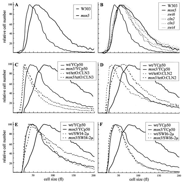
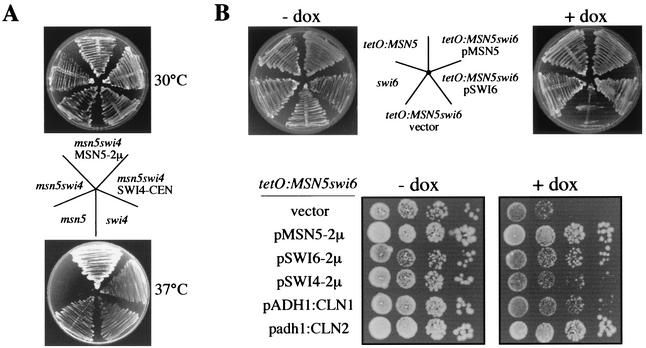
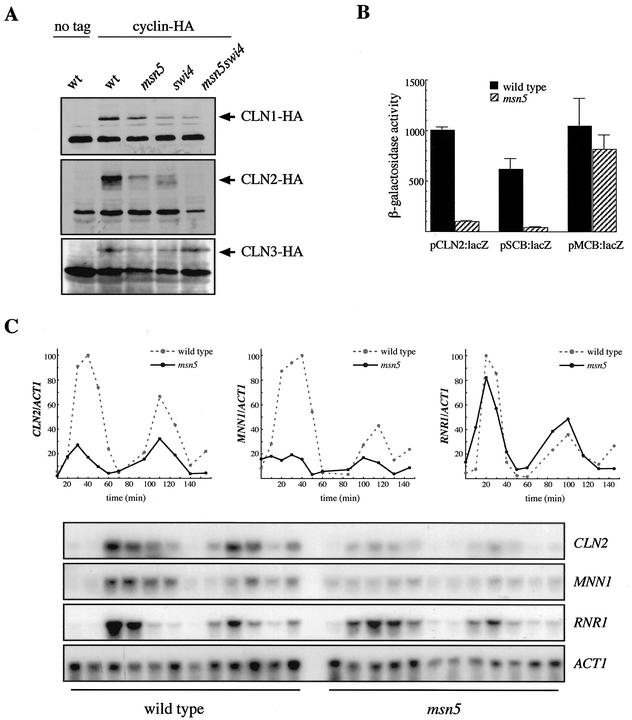
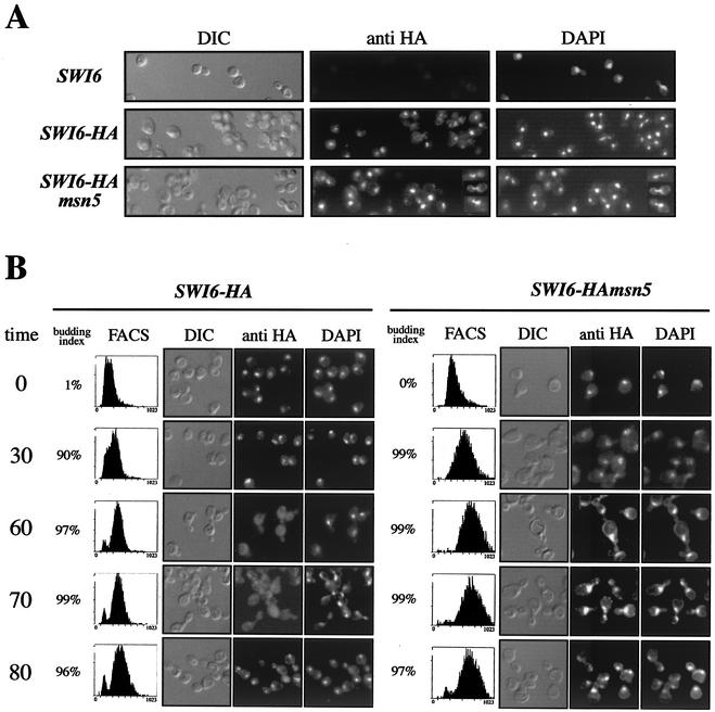
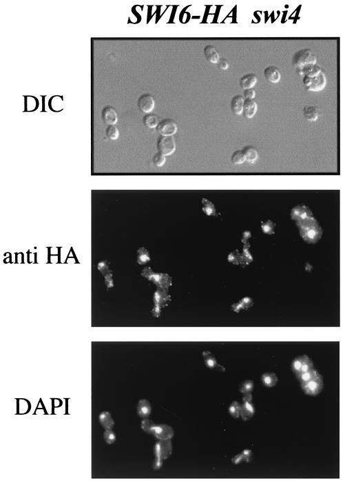
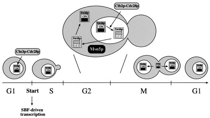
The control of the subcellular localization of cell cycle regulators has emerged as a crucial mechanism in the regulation of cell division. In the present work, we have characterized the function of the karyopherin Msn5p in the control of the cell cycle of Saccharomyces cerevisiae. Phenotypic analysis of the msn5 mutant revealed an increase in cell size and a functional interaction between Msn5p and the cell cycle transcription factor SBF (composed of the Swi4p and Swi6p proteins), indicating that Msn5p is involved in Start control. In fact, we have shown that the level of Cln2p protein is drastically reduced in an msn5 mutant. The effect on CLN2 expression is mediated at a transcriptional level, Msn5p being necessary for proper SBF-dependent transcription. On the contrary, loss of MSN5 has no effect on the closely related transcription factor MBF (composed of the Mbp1p and Swi6p proteins). Regulation of SBF by Msn5p is exerted by control of the localization of the regulatory subunit Swi6p. Swi6p shuttles between the nucleus and the cytoplasm during the cell cycle, and we have found that Msn5p is required for Swi6p export from the nucleus during the G(2)-M phase. What is more important, we have demonstrated that export of Swi6p to the cytoplasm is required for SBF activity, providing evidence for a functional switch of Swi6p linked to its nucleocytoplasmic shuttling during the cell cycle.


Figures

FIG. 1.
FIG. 1.
Analysis of the cell size in an msn5 mutant strain. (A and B) The sizes of the cells in exponentially growing cultures on YPD medium of wild-type (W303-1a), msn5 (JCY196), swi6 (JCY220), swi4 (JCY167), cln2 (JCY296), and cln3 (MT244) strains was determined in a Coulter cell counter. (C through F) Wild-type (wt) and msn5 mutant strains transformed with a control vector (YCp50), plasmids overexpressing CLN3 (ptetO:CLN3) or CLN2 (ptetO:CLN2), or multicopy plasmids containing the SWI6 or SWI4 gene were grown on synthetic medium, and cell sizes were determined. Graph are for the moving averages from at least four independent cultures of each of the strains.
FIG. 2.
FIG. 2.
Synthetic lethality between mutation in Msn5p and SBF components. (A) The swi4ts (BAM1-11A), msn5 (JCY196), and msn5 swi4ts (JCY047) strains, and the last transformed with a plasmid containing either the SWI4 or MSN5 gene, were streaked onto a YPD plate. Plates were incubated for 3 days at 30 or 37°C. (B) Upper panel, the swi6 (JCY220), tetO7:MSN5 (JCY324), and the tetO7:MSN5 swi6 (JCY335) strains, and the last transformed with a plasmid containing either the SWI6 or MSN5 gene, were streaked onto YPD plates with or without doxycycline and incubated at 28°C for 3 days. Lower panel, 10-fold serial dilutions from exponentially growing cultures of the tetO7:MSN5 swi6 strain transformed with a control vector, a multicopy plasmid containing the MSN5, SWI6, or SWI4 gene, a plasmid with the CLN1 gene under the control of the strong S. cerevisiae ADH1 promoter, or a plasmid bearing the CLN2 gene under the control of the mild Schizosaccharomyces pombe adh promoter were spotted onto medium with or without doxycycline (dox) and incubated at 30°C for 3 days.
FIG. 3.
FIG. 3.
Analysis of the regulation of gene expression by Msn5p. (A) Cln protein levels in extracts from wild-type (wt; W3031-a), msn5 (JCY196), swi4ts (BAM1-11A), and msn5 swi4ts (JCY047) strains bearing a centromeric plasmid containing the HA-tagged CLN1 (pCM137), CLN2 (pCM239), or CLN3 (pCM194) gene were examined by Western analysis. Control extracts from the wild-type strain transformed with an empty vector were included. A nonspecific band that cross-reacts with the 12CA5 antibody is shown as the loading control. (B) β-Galactosidase activity in extracts from wild-type (W303-1A) and msn5 (JCY179) strains transformed with pLGL-derived plasmids containing the lacZ reporter gene under the control of a fragment from the CLN2 promoter (pCLN2:lacZ), a fragment from the HO promoter with three SCB elements (pSCB:lacZ), or a synthetic oligonucleotide with three MCB elements (pMCB:lacZ) is shown. Values are derived from more than five extracts from at least three independent transformants. (C) Expression of the CLN2, MNN1, and RNR1 genes in α-factor-synchronized cultures of wild-type (W303-1A) and msn5 (JCY196) strains was studied by Northern analysis. Samples were collected at 0, 10, 20, 30, 40, 50, 60, 85, 100, 115, 130, and 145 min after release. ACT1 is shown as the loading control. Plots represent the amounts of CLN2, MNN1, and RNR1 mRNAs relative to that of ACT1 mRNA.
FIG. 4.
FIG. 4.
Control of Swi6p localization by Msn5p. (A) Cells from asynchronous cultures of wild-type (JCY114) and msn5 (JCY287) strains expressing a HA-epitope-tagged Swi6p protein were assayed by indirect immunofluorescence as described in Materials and Methods. Differential interference-contrast (DIC) images, the Swi6p-HA indirect-fluorescence signal (anti-HA), and DAPI staining of DNA are shown. The first row corresponds to a control of the untagged wild-type strain. Several mitotic cells demonstrating the nuclear localization of Swi6p during mitosis in the msn5 mutant strain (see text) are included to the right of the corresponding pictures. (B) Exponentially growing cultures of the same strains were arrested with α-factor for 3 h and released. Samples were taken during the arrest and at 30, 60, 70, and 80 min after release and were analyzed for budding index, DNA content (by FACS analysis), and Swi6p localization (by indirect immunofluorescence). Representative cells are shown.
FIG. 5.
FIG. 5.
Two-hydrid interaction assay between Msn5p and Swi6p. A plasmid expressing a fusion between Msn5p and the Gal4p DNA-binding domain (pBD-MSN5) or the corresponding empty vector (pBD) and a multicopy plasmid containing SWI6 (pSWI6-2μ) or the corresponding empty vector were introduced in the PJ69-4A strain and in JCY450 (swi4 mutant strain derived from PJ69-4A) in different combinations as indicated. The ability of the proteins to induce expression of a GAL7:lacZ gene was tested by measuring β-galactosidase activity in extracts from exponentially growing cells. Values are derived from more than eight extracts from at least three independent transformants.
FIG. 6.
FIG. 6.
Assay of the association of Swi6p with the CLN2 and RNR1 promoters. Cells from asynchronous cultures of wild-type (JCY114; lane SWI6-HA) and msn5 (JCY287; lane SWI6-HAmsn5) strains expressing a HA-epitope-tagged Swi6p protein were tested for Swi6p binding to the CLN2 and RNR1 promoters by chromatin immunoprecipitation assays. DNA samples were purified after cross-linking and immunoprecipitation and analyzed by PCR with primer pairs amplifying fragments from the CLN2, RNR1, MAT, or URA3 promoter. Control lanes show DNA amplified from different amounts of total DNA prior to purification (whole-cell extract [WCE]) and from the extract of a strain with untagged protein (W303-1a; lane SWI6)
FIG. 7.
FIG. 7.
Analysis of Swi4p protein level and localization in an msn5 mutant strain. (A) Swi4p protein levels in extracts from wild-type (JCY331; lane SWI4-HA) and msn5 (JCY333; lane SWI4-HAmsn5) strains expressing a HA-epitope-tagged Swi4p protein. Control extracts from the untagged wild-type strain were included. A nonspecific band labeled with an asterisk is shown as the loading control. (B) Cells from asynchronous cultures of the JCY331 (SWI4-HA) and JCY333 (SWI4-HAmsn5) strains were assayed by indirect immunofluorescence as described in the legend to Fig. 4. DIC, differential interference-contrast.
FIG. 8.
FIG. 8.
One cell cycle assay of the effect of blockage of nuclear export on Swi6p function. The JCY287 strain (SWI6-HAmsn5) transformed with the pGAL:MSN5 plasmid was grown on galactose medium and arrested by α-factor. Once blocked, culture was split and cells were transferred to YPGal or YPD medium containing α-factor. After two additional hours in the presence of α-factor, cells were released from the arrest, and after 40 min, α-factor was reinoculated to block progression in the next cycle. Samples were collected before the release and after the arrest in the next cycle (samples 1 and 2 for cells incubated on YPGal medium and samples 3 and 4 for cells grown on YPD medium). An aliquot of the cells transferred to YPD medium was maintained arrested during the whole experiment (sample 5). Samples were investigated for the subcellular localization of Swi6p by indirect immunofluorescence (as described in the legend to Fig. 4) and for association of Swi6p to the CLN2 promoter by chromatin immunoprecipitation (as described in the legend to Fig. 6). WCE, whole-cell extract; DIC, differential interference-contrast.
FIG. 9.
FIG. 9.
Effect of PKI NES-driven nuclear export on Swi6p function. The activity of a HA-epitope-tagged Swi6p protein fused to an active (Swi6pNES) or inactive (Swi6pnesi) version of the NES from the PKI protein was assayed in the wild type (JCY438 and JCY440 strains) and msn5 mutant (JCY442 and JCY444 strains) backgrounds. Subcellular localization of Swi6p (A), cell size (B), expression of the SCB:lacZ reporter gene (C), and binding of Swi6p to the CLN2 promoter (D) were analyzed as described previously. WCE, whole-cell extract; DIC, differential interference-contrast.
FIG. 10.
FIG. 10.
Subcellular localization of Swi6p in a swi4 mutant strain. Cells of a swi4 mutant strain expressing a HA-epitope-tagged Swi6p (JCY353) protein were fixed and assayed by indirect immunofluorescence as described in the legend to Fig. 4. DIC, differential interference-contrast.
FIG. 11.
FIG. 11.
Functional cycle of Swi6p. Different mechanisms affect Swi6p function and localization during the cell cycle. Swi6p is imported into the nucleus at the end of mitosis. As a part of the SBF transcription factor, it binds to the target promoter, although gene expression is not triggered until SBF is activated at Start by the Cln3p-Cdc28p kinase. This activation leads to progression through the cell cycle and subsequently to the accumulation of Clb-Cdc28p kinase activity. This kinase is responsible for the inactivation of SBF in the G2 phase, displacing it from the DNA. By this or another additional mechanism, Swi6p is targeted for being exported to the cytoplasm by the karyopherin Msn5p. Relocation of Swi6p to the cytoplasm enables a functional re-setting of Swi6p, rendering a protein able to activate transcription in the next cycle.

Similar articles

Cited by

References

    1. Alepuz, P. M., D. Matheos, K. W. Cunningham, and F. Estruch. 1999. The Saccharomyces cerevisiae RanGTP-binding protein Msn5p is involved in different signal transduction pathways. Genetics 153:1219-1231. - PMC - PubMed
    1. Amon, A., M. Tyers, B. Futcher, and K. Nasmyth. 1993. Mechanisms that help the yeast cell cycle clock tick: G2 cyclins transcriptionally activate G2 cyclins and repress G1 cyclins. Cell 74:993-1007. - PubMed
    1. Baetz, K., and B. Andrews. 1999. Regulation of cell cycle transcription factor Swi4 through auto-inhibition of DNA binding. Mol. Cell. Biol. 19:6729-6741. - PMC - PubMed
    1. Bartek, J., J. Bartkova, and J. Lukas. 1996. The retinoblastoma protein pathway and the restriction point. Curr. Opin. Cell Biol. 8:805-814. - PubMed
    1. Benton, B. K., A. H. Tinkelenberg, D. Jean, S. D. Plump, and F. R. Cross. 1993. Genetic analysis of Cln/Cdc28 regulation of cell morphogenesis in budding yeast. EMBO J. 12:5267-5275. - PMC - PubMed

Publication types

MeSH terms