. 2003 May;23(9):3247-52.
doi: 10.1128/MCB.23.9.3247-3252.2003.

Targeted disruption of the peptide transporter Pept2 gene in mice defines its physiological role in the kidney

Affiliations

Targeted disruption of the peptide transporter Pept2 gene in mice defines its physiological role in the kidney

Isabel Rubio-Aliaga et al. Mol Cell Biol. 2003 May.

Abstract

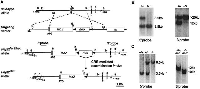
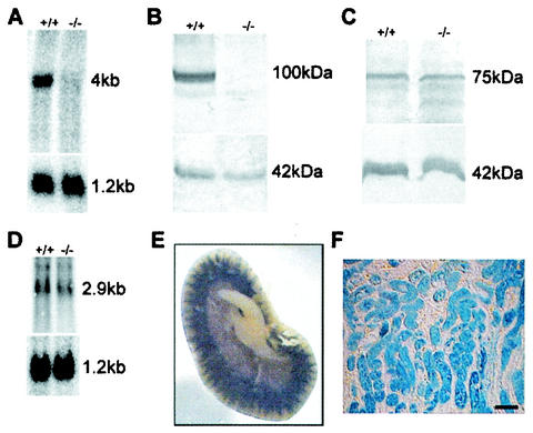
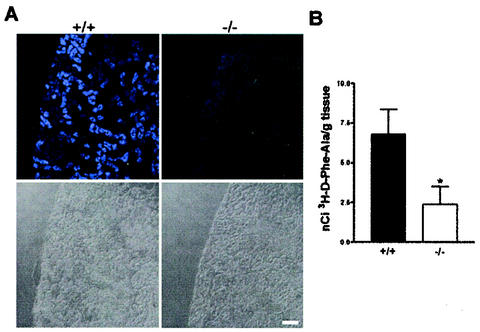
The peptide transporter PEPT2 mediates the cellular uptake of di- and tripeptides and selected drugs by proton-substrate cotransport across the plasma membrane. PEPT2 was functionally identified initially in the apical membrane of renal tubular cells but was later shown to be expressed in other tissues also. To investigate the physiological importance of PEPT2 and for a detailed analysis of the protein expression sites, we generated a Pept2 knockout mouse line in which the Pept2 gene was disrupted by insertion of a beta-galactosidase gene under the control of the PEPT2 promoter. The Pept2(-/-) mice showed no obvious phenotypic abnormalities but also no adaptive upregulation in the expression level of related genes in the kidney. The importance of PEPT2 in the reabsorption of filtered dipeptides was demonstrated in knockout animals by significantly reduced renal accumulation of a fluorophore-labeled and a radiolabeled dipeptide after in vivo administration of the tracers. This indicates that PEPT2 is the main system responsible for tubular reabsorption of peptide-bound amino acids, although this does not lead to major changes in renal excretion of protein or free amino acids.


Figures

FIG. 1.
FIG. 1.
Targeted disruption of the mouse Pept2 gene. (A) Schematic representation of the genomic Pept2 locus, the targeting construct, and the different mutated alleles. Homologous recombination of the targeting vector introduces the lacZ gene downstream of the Pept2 promoter, deleting the coding region of the first exon and the loxP-flanked neo gene. Cre-mediated recombination in vivo produces the excision of the neomycin resistance gene. Black boxes represent the coding exons, whereas the shaded box represents the noncoding region of exon 1. loxP sites are indicated as solid triangles. Dashed lines show regions of identity between the locus and the targeting vector. Grey bars indicate the 5′ internal and 3′ external probes used for the detection of the targeted allele by Southern blot analysis. tk, selectable thymidine kinase gene. B, BamHI; E, EcoRI; Sc, SacI; Xh, XhoI. (B) Southern blot analysis of genomic DNA from targeted ES cells. After digestion with EcoRI and probing with the 5′ internal probe, a 6.5-kb restriction fragment is diagnostic for the mutated allele (−/−), in contrast to the 3.5-kb fragment generated from the wild-type allele (+/+) (left). With the 3′ external probe and after digestion with BamHI, a 12-kb fragment indicates the mutated allele, whereas a fragment greater than 20 kb represents the wild-type allele (right). (C) Southern blot analysis of genomic DNA from mouse tail biopsy samples by digestion with EcoRI and hybridization with the 5′ probe (left). After in vivo Cre recombination digestion with BamHI and hybridization with the 3′ probe, a 10-kb instead of a 12-kb fragment indicates the excision of the neo gene in the mutant allele (right).
FIG. 2.
FIG. 2.
Mutated Pept2 allele is a null allele, and no compensatory upregulation is found in related genes. (A) Northern blot analysis with RNA isolated from the kidneys of wild-type (+/+) and Pept2 knockout (−/−) mice. The 4-kb fragment corresponding to the PEPT2 mRNA was not detectable in the RNA from the Pept2 knockout mice. The blot was stripped and rehybridized with a glyceraldehyde-3-phosphate dehydrogenase probe as a loading and quality control (1.2-kb fragment). (B) Western blot analysis showing the absence of the PEPT2 protein in the kidney. The 100-kDa protein corresponding to the PEPT2 protein was not detectable in the knockout mice. The blot was probed with an antibody directed against actin as a quality and quantity control (42-kDa fragment). (C) Western blot analysis showing no increase in the PEPT1 protein level in the kidney. No differences were detected in the intensity of the 75-kDa band, which corresponds to the PEPT1 protein. The blot was probed with an antibody directed against actin as a quality and quantity control (42-kDa fragment). (D) Northern blot analysis showing no increase in the PHT1 mRNA level in the kidney. No differences were detected in the intensity of the 2.9-kb band which corresponds to the PHT1 RNA. The blot was striped and rehybridized with a glyceraldehyde-3-phosphate dehydrogenase probe as a loading and quality control (1.2-kb fragment). (E) Tissue overview after LacZ staining of kidney from adult Pept2−/− mice and slices (F). Bar, 80 μm.
FIG. 3.
FIG. 3.
Reduced renal accumulation of a fluorophore-conjugated dipeptide (A) and a radiolabeled-dipeptide (B) in Pept2 knockout mice. (A) In kidney slices obtained from wild-type (+/+) mice after intravenous injection of d-Ala-Lys-AMCA, fluorescence was detectable in selected tubular structures of the cortex and outer medulla, whereas in slices from the Pept2 knockout (−/−) mice, only very weak fluorescence was detectable in the outer cortex. Bottom, light microscopy pictures of the corresponding kidney sections. Bar, 80 μm. (B) In kidney homogenates prepared from Pept2 knockout mice (−/−) and wild-type animals (+/+), accumulation of radioactivity after intraperitoneal administration of d-[3H]Phe-Ala was significantly (*, P < 0.05) reduced.

Similar articles

Cited by

References

    1. Adibi, S. A. 1997. Renal assimilation of oligopeptides: physiological mechanisms and metabolic importance. Am. J. Physiol. 272:E723-E736. - PubMed
    1. Botka, C. W., T. W. Wittig, R. C. Graul, C. U. Nielsen, K. Higaka, G. L. Amidon, and W. Sadee. 2000. Human proton/oligopeptide transporter (POT) genes: identification of putative human genes with bioinformatics. AAPS Pharm Sci. 2:E16. - PMC - PubMed
    1. Broccoli, V., E. Boncinelli, and W. Wurst. 1999. The caudal limit of Otx2 expression positions the isthmic organizer. Nature 401:164-168. - PubMed
    1. Daniel, H., and M. Herget. 1997. Cellular and molecular mechanisms of renal peptide transport. Am. J. Physiol. 273:F1-F8. - PubMed
    1. Daniel, H., E. L. Morse, and S. A. Adibi. 1991. The high and low affinity transport systems for dipeptides in kidney brush border membrane respond differently to alterations in pH gradient and membrane potential. J. Biol. Chem. 266:19917-19924. - PubMed

Publication types

LinkOut - more resources