. 2003 May;23(9):3305-19.
doi: 10.1128/MCB.23.9.3305-3319.2003.

Two Drosophila Ada2 homologues function in different multiprotein complexes

Affiliations

Two Drosophila Ada2 homologues function in different multiprotein complexes

Thomas Kusch et al. Mol Cell Biol. 2003 May.

Abstract

The reversible acetylation of the N-terminal tails of histones is crucial for transcription, DNA repair, and replication. The enzymatic reaction is catalyzed by large multiprotein complexes, of which the best characterized are the Gcn5-containing N-acetyltransferase (GNAT) complexes. GNAT complexes from yeast to humans share several conserved subunits, such as Ada2, Ada3, Spt3, and Tra1/TRRAP. We have characterized these factors in Drosophila and found that the flies have two distinct Ada2 variants (dAda2a and dAda2b). Using a combination of biochemical and cell biological approaches we demonstrate that only one of the two Drosophila Ada2 homologues, dAda2b, is a component of Spt-Ada-Gcn5-acetyltransferase (SAGA) complexes. The other Ada2 variant, dAda2a, can associate with dGcn5 but is not incorporated into dSAGA-type complexes. This is the first example of a complex-specific association of the Ada-type transcriptional adapter proteins with GNATs. In addition, dAda2a is part of Gcn5-independent complexes, which are concentrated at transcriptionally active regions on polytene chromosomes. This implicates novel functions for dAda2a in transcription. Humans and mice also possess two Ada2 variants with high homology to dAda2a and dAda2b, respectively. This suggests that the mammalian and fly homologues of the transcriptional adapter Ada2 form two functionally distinct subgroups with unique characteristics.


Figures
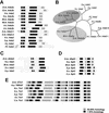
FIG. 1.
FIG. 1.
Homology plots of the Drosophila GNAT subunits dAda2a and dAda2b (A), dAda3 (C), dSpt3 (D), and dTra1 (E). (B) Unrooted phylogenetic tree generated with the CLUSTAL W (47) and PHYLIP (21) programs of the Ada2 homologues from various species (bootstrap value, 100; 15 repetitions). The distinct Ada2 variants from human, mouse, and Drosophila lie within the same branch (shaded ovals). Abbreviations: D.m., D. melanogaster; H.s., Homo sapiens; M.m., Mus musculus; A.t., Arabidopsis thaliana; Z.m., Zea mais; S.c., Saccharomyces cerevisiae; S.p., Schizosaccharomyces pombe; P.f., Plasmodium falciparum; C.o., Clavispora opuntiae; C.e., C. elegans; H.j., Hypocrea jecorina.
FIG. 2.
FIG. 2.
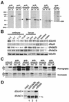
Characterization of the Drosophila GNAT subunits dAda2a, dAda2b, dAda3, dSpt3, and dTra1. (A) The antisera (is, immune serum) recognize proteins with the predicted molecular masses of dGcn5, dAda2b, dAda2a, dAda3, dSpt3, and dTra1 (arrowheads). Each lane contains 25 μg of nuclear extract. pre, preimmunization bleeds. Crude antisera against dAda2a and Spt3 detect a cross-reactive band of similar molecular mass, which is not recognized by affinity-purified antibodies (ap). (B) Developmental expression of dGcn5, dSpt3, dAda2a, and dAda2b. Lysates from Oregon R animals were electrophoresed on an SDS-10% polyacrylamide gel and analyzed by Western blotting. Embryonic lysates: 0-3 (blastoderm), 0-7 (gastrulation), 7-13 (germ band retraction), and 13-24 (differentiation). Larval stages: first, second, and third instars. Antibodies against tubulin served as loading controls. (C) HAT activity of the immunoprecipitates from 300 μg of S2 nuclear extract on nucleosomes (nuc) and core histones (ch), respectively. dAda3, dAda2a, dAda2b, and dSpt3 are associated with a dGcn5-specific HAT activity. Bacterially expressed rdGcn5 was used as a control. Top, fluorogram; bottom, photography of the Coomassie brilliant blue-stained SDS-polyacrylamide gel. (D) Western blot probed for dAda3 and dGcn5 (arrowheads). Input, 30 μg of nuclear extract; lane 1, immunoprecipitates from anti-dAda2b antibodies; lane 2, immunoprecipitates from anti-dAda2a; lane 3, immunoprecipitates from anti-dSpt3. hc, IgG heavy chain.
FIG. 3.
FIG. 3.
dAda2b, but not dAda2a, associates with dmTaf5, dmTaf9, dmTaf10, dSpt3, and dTra1. Affinity-purified antibodies were conjugated to protein A-Sepharose beads and were used in immunoprecipitation assays of 300 μg of nuclear extract from S2 cells. Input, 30 μg of nuclear extract. (A) Western blots immunoprobed for dSpt3 (left) and dTra1 (right). Lane 1, immunoprecipitates from anti-dAda2a (αdAda2a); lane 2, immunoprecipitates from anti-dAda2b; lane 3, immunoprecipitates from anti-dGcn5; lane 4, immunoprecipitates from anti-dAda3; lane 5, immunoprecipitates from anti-dAda2a; lane 6, immunoprecipitates from anti-dAda2b. (B) Western blots probed for dAda2a (left) and dAda2b (right). Lane 1, immunoprecipitates from anti-dSpt3; lane 2, immunoprecipitates from anti-dAda2b; lane 3, immunoprecipitates from anti-dGcn5; lane 4, immunoprecipitates from anti-dAda3; lane 5, immunoprecipitates from anti-dAda2a; lane 6, immunoprecipitates from anti-dSpt3; lane 7, immunoprecipitates from anti-dGcn5; lane 8, immunoprecipitates from anti-dAda3. hc, IgG heavy chain. (C) Western blots immunolabeled with antibodies against dmTaf5, dmTaf9, and dmTaf10. Lane 1, immunoprecipitates from anti-dAda2b; lane 2, immunoprecipitates from anti-dSpt3; lane 3, immunoprecipitates from anti-dAda3; lane 4, immunoprecipitates from anti-dAda2a.
FIG. 4.
FIG. 4.
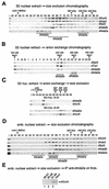
dAda2b associates with dSAGA-specific subunits in biochemical fractionation assays. (A) Elution profiles of dGcn5, dAda3, dSpt3, dAda2b, and dAda2a from a Sephacryl S400 gel filtration column; 5 mg of S2 nuclear extract was separated on the column, and 15 μl of each fraction was subjected to SDS-PAGE, followed by Western blotting (fraction numbers indicated at the top). dGcn5 and dAda3 coelute in a range from ∼2.2 MDa to 300 kDa. dAda2b coelutes with dSpt3 in a range of ∼2 MDa to 800 kDa. dAda2a elutes in two peaks around 2.5 MDa and 440 kDa. i, input (30 μg of embryonic nuclear extract). (B) dSpt3, and dTra1 cofractionate with dAda2b, but not dAda2a, in anion-exchange chromatography. Bound proteins were eluted in a linear gradient of 100 to 450 mM sodium chloride from a 1-ml MonoQ anion-exchange column. The salt concentrations of the peak fractions are indicated above the blot. Fifteen microliters of each fraction was subjected to SDS-PAGE, followed by Western blotting (fraction numbers indicated at the top; f1 to f3, flowthrough). (C) MonoQ (MQ) fractions (frcts.) 16 to 20 (top) and 24 to 28 (bottom) of the anion-exchange chromatography were concentrated and subjected to size exclusion chromatography. dAda2b and dGcn5 coelute in a range of ∼1.8 MDa. dAda2a elutes in fractions of >2 MDa and ∼450 kDa. dGcn5 is detectable only in fractions containing the smaller dAda2a complex. nuc., nuclear. (D) Gel filtration analysis of nuclear extract from 0- to 12-h-old embryos as described for panel A. A third dAda2a peak is observed in a molecular-mass range of ∼1.8 MDa. (E) Pooled dAda2a peak fractions from embryonic extracts (panel D) were subjected to immunoprecipitation assays with affinity-purified anti-dAda2a antibodies. The immunoprecipitates (IP) were separated by SDS-PAGE and were probed for dGcn5. Lane 1, fractions 58 to 61; lane 2, fractions 65 to 68; lane 3, fractions 81 to 84.
FIG. 5.
FIG. 5.
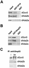
VP16 and Dmp53 interact with dAda2b-containing GNAT complexes. (A) GST pull-down assays were performed with GST-VP16 or GST alone bound to GSH-Sepharose beads and 300 μg of S2 nuclear extract. The precipitates were resolved by SDS-PAGE and transferred onto nitrocellulose membranes for immunodetection assays with antisera against dGcn5, dAda2b, and dAda2a. Input, 30 μg of nuclear extract. (B) GST pull-down assays using a GST-Dmp53 fusion protein. (C) Coimmunoprecipitation assays were performed with anti-Dmp53 antibodies conjugated to protein A-Sepharose and 300 μg of S2 nuclear extract. The immunoprecipitated (IP) proteins were separated by SDS-PAGE prior to Western blotting analyses using antisera against dGcn5, dAda2b, and dAda2a. i, input; be, beads.
FIG. 6.
FIG. 6.
dAda3 and dGcn5 colocalize on polytene chromosomes from third-instar larvae. Polytene chromosome spreads were double labeled with affinity-purified antibodies against dAda3 (green) and dGcn5 (red). (A) Staining with rabbit anti-dAda3 antibodies. (B) Staining with rat anti-dGcn5 antibodies. (C) Overlap of green and red stains appears in the merge of panels A and B as yellow, yellow-green, and orange. dGcn5 and dAda3 show almost complete overlap. (D) Magnification of the boxed region in panel C. DNA was counterstained with DAPI, which preferentially labels heterochromatin. Note that dAda3 and dGcn5 mainly localize to DAPI-negative euchromatic regions.
FIG. 7.
FIG. 7.
Localization studies of dAda2b and dAda2a on polytene chromosomes from third-instar larvae. Polytene chromosome spreads were double-labeled with the indicated affinity-purified antibodies. The pictures were taken at different magnifications; the bars correspond to 20 μm. (A to D) Merged red and green channels. (A) Costaining using rabbit anti-dAda3 antibodies (red) and rat anti-dAda2b antibodies (green). The arrowheads indicate selected regions exclusively staining for dAda3. (B) Costaining with rabbit anti-dAda2a (red) and rat anti-dAda2b (green). The arrowheads indicate selected regions of signal overlap. The arrows indicate puffs strongly staining for dAda2a. (C) Costaining with rat anti-dAda2a (red) and rabbit anti-dAda3 (green). The arrowheads indicate selected regions of signal overlap. The arrows indicate puffs strongly staining for dAda2a. (D) Costaining with rabbit anti-dAda2a (red) and rat anti-dGcn5 (green). The arrowheads indicate selected regions of overlap. The arrows indicate puffs strongly staining for dAda2a. (E) Magnification of the boxed region in panel C. DNA was stained with DAPI. dAda2a strongly stains decondensed euchromatic regions (compare the white bars). dAda3 and dAda2a colocalize in less condensed euchromatic regions. (F) dAda2a and phosphorylated RNA polymerase II colocalize on polytene chromosomes from third-instar larvae. Top, staining with rabbit anti-dAda2a antibodies; middle, staining with mouse anti-phosphorylated C-terminal domain of RNA polymerase II antibodies; bottom, overlap of green and red stains appears in the merge of panels A and B as yellow, yellow-green, and orange.
FIG. 7.
FIG. 7.
Localization studies of dAda2b and dAda2a on polytene chromosomes from third-instar larvae. Polytene chromosome spreads were double-labeled with the indicated affinity-purified antibodies. The pictures were taken at different magnifications; the bars correspond to 20 μm. (A to D) Merged red and green channels. (A) Costaining using rabbit anti-dAda3 antibodies (red) and rat anti-dAda2b antibodies (green). The arrowheads indicate selected regions exclusively staining for dAda3. (B) Costaining with rabbit anti-dAda2a (red) and rat anti-dAda2b (green). The arrowheads indicate selected regions of signal overlap. The arrows indicate puffs strongly staining for dAda2a. (C) Costaining with rat anti-dAda2a (red) and rabbit anti-dAda3 (green). The arrowheads indicate selected regions of signal overlap. The arrows indicate puffs strongly staining for dAda2a. (D) Costaining with rabbit anti-dAda2a (red) and rat anti-dGcn5 (green). The arrowheads indicate selected regions of overlap. The arrows indicate puffs strongly staining for dAda2a. (E) Magnification of the boxed region in panel C. DNA was stained with DAPI. dAda2a strongly stains decondensed euchromatic regions (compare the white bars). dAda3 and dAda2a colocalize in less condensed euchromatic regions. (F) dAda2a and phosphorylated RNA polymerase II colocalize on polytene chromosomes from third-instar larvae. Top, staining with rabbit anti-dAda2a antibodies; middle, staining with mouse anti-phosphorylated C-terminal domain of RNA polymerase II antibodies; bottom, overlap of green and red stains appears in the merge of panels A and B as yellow, yellow-green, and orange.

Similar articles

Cited by

References

    1. Aasland, R., A. F. Stewart, and T. Gibson. 1996. The SANT domain: a putative DNA-binding domain in the SWI-SNF and ADA complexes, the transcriptional co-repressor N-CoR and TFIIIB. Trends Biochem. Sci. 21:87-88. - PubMed
    1. Allard, S., R. T. Utley, J. Savard, A. Clarke, P. Grant, C. J. Brandl, L. Pillus, J. L. Workman, and J. Cote. 1999. NuA4, an essential transcription adaptor/histone H4 acetyltransferase complex containing Esa1p and the ATM-related cofactor Tra1p. EMBO J. 18:5108-5119. - PMC - PubMed
    1. Altschul, S. F., W. Gish, W. Miller, E. W. Myers, and D. J. Lipman. 1990. Basic local alignment search tool. J. Mol. Biol. 215:403-410. - PubMed
    1. Balasubramanian, R., M. G. Pray-Grant, W. Selleck, P. A. Grant, and S. Tan. 2002. Role of the Ada2 and Ada3 transcriptional coactivators in histone acetylation. J. Biol. Chem. 277:7989-7995. - PubMed
    1. Berger, S. L., B. Pina, N. Silverman, G. A. Marcus, J. Agapite, J. L. Regier, S. J. Triezenberg, and L. Guarente. 1992. Genetic isolation of ADA2: a potential transcriptional adaptor required for function of certain acidic activation domains. Cell 70:251-265. - PubMed

MeSH terms