. 2003 May;23(9):3320-8.
doi: 10.1128/MCB.23.9.3320-3328.2003.

Dimerization of MLH1 and PMS2 limits nuclear localization of MutLalpha

Affiliations

Dimerization of MLH1 and PMS2 limits nuclear localization of MutLalpha

Xiaosheng Wu et al. Mol Cell Biol. 2003 May.

Abstract

DNA mismatch repair maintains genomic stability by detecting and correcting mispaired DNA sequences and by signaling cell death when DNA repair fails. The mechanism by which mismatch repair coordinates DNA damage and repair with cell survival or death is not understood, but it suggests the need for regulation. Since the functions of mismatch repair are initiated in the nucleus, we asked whether nuclear transport of MLH1 and PMS2 is limiting for the nuclear localization of MutLalpha (the MLH1-PMS2 dimer). We found that MLH1 and PMS2 have functional nuclear localization signals (NLS) and nuclear export sequences, yet nuclear import depended on their C-terminal dimerization to form MutLalpha. Our studies are consistent with the idea that dimerization of MLH1 and PMS2 regulates nuclear import by unmasking the NLS. Limited nuclear localization of MutLalpha may thus represent a novel mechanism by which cells fine-tune mismatch repair functions. This mechanism may have implications in the pathogenesis of hereditary non-polyposis colon cancer.


Figures

FIG. 1.
FIG. 1.
MLH1 and PMS2 have putative NLS and NES and localize in the nucleus. (A) Domain structure of mouse MLH1 and PMS2. C-terminal dimerization domains were identified as described by Guerrette et al. (16), and ATP binding domains were identified as described by Ban and Yang (2). Putative NLS and NES were identified by homology with known functional sequences from simian virus 40 large T antigen (32), c-myc (20), human ribosomal protein S6 (39), human immunodeficiency virus rev protein (42), human cyclic AMP-dependent protein kinase inhibitor (42), and p53 (46). The alignment identified three putative NLS in the PMS2 and one in the MLH1. In addition, three putative NES sequences were identified in PMS2 and MLH1. Mouse and human MLH1 and PMS2 NLS and NES were identical. (B to E) Confocal microscopy showing the subcellular localization of endogenous MLH1 (B) and PMS2 (D) in HeLa cells. 4′,6′-Diamidino-2-phenylindole (DAPI) nuclear staining is shown in panels C and E. Images are representative of 99% of the cells examined (496 out of 500).
FIG. 2.
FIG. 2.
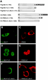
MLH1 and PMS2 possess functional NLS. (A) MLH1 and PMS2 full-length and mutant constructs. An F- or H-marked box identifies the Flag or the HA tag cloned in frame 5′ to the coding sequence of MLH1 or PMS2, respectively. The NLS are identified by black-filled rectangles, and the NES are identified by gray-filled rectangles. The hatched areas represent the C-dimerization domain of MLH1 or PMS2, respectively. (B to G) Intracellular localization of MLH1 (green) or PMS2 (red) in HCT 116 cells transfected with Flag-MLH1 (B), Flag-MLH1ΔC (D), Flag-MLH1ΔC-ΔNLS (F), HA-PMS2 (C), HA-PMS2ΔC (E), or HA-PMS2ΔC-ΔNLS (G). MLH1 or PMS2 was detected with anti-Flag (B, D, and F) or anti-HA (C, E, and G) antibodies, respectively, as indicated. Images C to G are representative of 487 (97%), 382 (76%), 474 (95%), 481 (96%), and 477 (95%) cells out of 500 transfected cells examined. (B) Flag-MLH1 was exclusively localized to the cytoplasm in 255 (51%) cells and is partitioned between the cytoplasm and the nucleus in 245 (49%) cells out of 500 transfectants examined. Confocal images were collected and processed by using the same settings, as explained in Materials and Methods.
FIG. 3.
FIG. 3.
MLH1 and PMS2 possess functional NES. (A) MLH1-NES (MNES-GrG), PMS2-NES (PNES-GrG), and control (GrG) reporter constructs. GrG is an abbreviation for glucocorticoid receptor (Gr)-GFP (G) fusion, M refers to MLH1, and P refers to PMS2. All constructs were cloned into the pUHD10S vector. (B to J) Intracellular localization of GFP in HtTA-expressing cells transfected with MNES-GrG (B, E, and H), PNES-GrG (C, F, and I), or control construct GrG (D, G, and J). Panels B, C, and D show transfectants not treated with dexamethasone (DEX); panels E, F, and G represent transfectants treated with 1 μM dexamethasone for 30 min; panels H, I, and J represent cells treated with dexamethasone, followed by removal for two hours, in the presence of 30 μg of cycloheximide/ml. Confocal images were representative of at least 85% of 500 transfected cells examined and were collected and processed by using the settings explained in Materials and Methods.
FIG. 4.
FIG. 4.
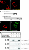
MLH1 and PMS2 nuclear import is impaired by their C-terminal domains. (A and B) Intracellular localization of HA-PMS2 expressed in HCT 116 cells with (B) or without (A) 20.0 ng of leptomycin B (LMB)/ml for 2 h prior to immunostaining. PMS2 was detected with an anti-HA antibody. (C) MLH1/PMS2 and PMS2/MLH1 chimeric constructs were obtained by replacing the C-terminal domains of MLH1 and PMS2 with each other. (D and E) Intracellular localization of HA-PMS2/MLH1 (D) or Flag-MLH1/PMS2 (E) fusion proteins in transfected HCT 116 cells. The HA or Flag tags were detected with anti-HA (D) or anti-Flag (E) antibodies, as indicated in the photographs. The confocal images were representative of at least 95% of 500 transfected cells analyzed. (F) Western blot (WB) analysis of immunoprecipitation (IP) of MLH1/PMS2 and PMS2/MLH1 chimeric proteins in total cell lysates of HCT 116 cells (lane 1) or HCT 116 cells transfected with HA-PMS2/MLH1 (lanes 2 and 4) or Flag-MLH1/PMS2 (lane 3 and 4). Whole-cell lysates were immunoprecipitated with anti-Flag or anti-HA antibodies, as indicated. Precipitated proteins were resolved on SDS-8% PAGE and were blotted onto PVDF membranes, followed by probing with anti-Flag or anti-HA antibodies, as indicated. Bands were visualized with the ECL detection kit.
FIG. 5.
FIG. 5.
MLH1 and PMS2 are imported into the nucleus as a C-terminal MutLα heterodimer. (A) MLH1 and PMS2 full-length or deletion mutants were marked as described in the legend to Fig. 2. (B to G) Intracellular localization of MLH1 (green) or PMS2 (red) in HCT 116 cells cotransfected with HA-PMS2 and Flag-MLH1 (B and C), Flag-MLH1ΔC (D and E), or Flag-MLH1ΔN (F and G). MLH1 or PMS2 was detected with anti-Flag (B, D, and F) or anti-HA (C, E, and G) antibodies. Images reflected 476 (95%) cells in panels B and C, 479 (96%) cells in panels D and E, and 485 (97%) cells in panels F and G, out of 500 transfected cells examined. Images were collected and processed by using the same settings, as explained in Materials and Methods. (H) Western blot (WB) analysis of immunoprecipitation (IP) of MLH1 and PMS2 in total cell lysates of HCT 116 cells (lane 1) or HCT 116 cells transfected with full-length HA-PMS2 (lanes 2 to 4), Flag-MLH1 (lane 2), Flag-MLH1ΔC (lane 3), or Flag-MLH1ΔN (lane 4). Whole-cell lysates were immunoprecipitated with anti-Flag or anti-HA antibodies, as indicated. Precipitated proteins were resolved on SDS-8% PAGE and were blotted onto PVDF membranes, followed by probing with anti-Flag or anti-HA antibodies, as indicated. Bands were visualized with the ECL detection kit. Ig, immunoglobulin.
FIG. 5.
FIG. 5.
MLH1 and PMS2 are imported into the nucleus as a C-terminal MutLα heterodimer. (A) MLH1 and PMS2 full-length or deletion mutants were marked as described in the legend to Fig. 2. (B to G) Intracellular localization of MLH1 (green) or PMS2 (red) in HCT 116 cells cotransfected with HA-PMS2 and Flag-MLH1 (B and C), Flag-MLH1ΔC (D and E), or Flag-MLH1ΔN (F and G). MLH1 or PMS2 was detected with anti-Flag (B, D, and F) or anti-HA (C, E, and G) antibodies. Images reflected 476 (95%) cells in panels B and C, 479 (96%) cells in panels D and E, and 485 (97%) cells in panels F and G, out of 500 transfected cells examined. Images were collected and processed by using the same settings, as explained in Materials and Methods. (H) Western blot (WB) analysis of immunoprecipitation (IP) of MLH1 and PMS2 in total cell lysates of HCT 116 cells (lane 1) or HCT 116 cells transfected with full-length HA-PMS2 (lanes 2 to 4), Flag-MLH1 (lane 2), Flag-MLH1ΔC (lane 3), or Flag-MLH1ΔN (lane 4). Whole-cell lysates were immunoprecipitated with anti-Flag or anti-HA antibodies, as indicated. Precipitated proteins were resolved on SDS-8% PAGE and were blotted onto PVDF membranes, followed by probing with anti-Flag or anti-HA antibodies, as indicated. Bands were visualized with the ECL detection kit. Ig, immunoglobulin.
FIG. 6.
FIG. 6.
Formation and nuclear import of transspecies MutLα. (A) Western blot analysis of immunoprecipitation of Flag-MLH1 and HA-PMS2 in whole-cell lysates of HtTA cells nontransfected (lane 1), transfected with HA-PMS2 alone (lane 2), or transfected with HA-PMS2 and Flag-MLH1 (lane 3). Immunoprecipitation (IP) was with anti-Flag or anti-HA antibodies, as indicated. Precipitated proteins were resolved on SDS-6% PAGE and blotted onto PVDF membranes (WB), followed by probing with anti-Flag, anti-HA, or anti-MLH1 antibodies as indicated. Bands were visualized with the ECL detection kit. (B) Intracellular localization of mouse MLH1 in HeLa cells transfected with Flag-MLH1. Mouse MLH1 protein was detected with anti-Flag antibody. Image represents more than 95% of 500 transfectants examined.
FIG. 7.
FIG. 7.
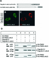
NLS of MLH1 and PMS2 are required for nuclear localization. (A) Schematic representation of NLS MLH1 and PMS2 deletion mutant constructs Flag-MLH1-ΔNLS and HA-PMS2-ΔNLS, respectively. (B and C) Intracellular localization of MLH1 (green) or PMS2 (red) mutants in HCT 116 cells cotransfected with Flag-MLH1-ΔNLS and HA-PMS2-ΔNLS. MLH1 or PMS2 was detected with anti-Flag (B) or anti-HA (C) antibodies. Images B and C represent 439 (88%) of 500 transfected cells examined and were collected and processed by using the same settings, as explained in Materials and Methods. (D) Western blot analysis of MLH1 and PMS2 immunoprecipitated from nontransfected HCT 116 cells (lane 2), cells transfected with full-length HA-PMS2 (lanes 1 and 4) and Flag-MLH1 (lane 1) or Flag-MLH1-ΔNLS (lane 4), and cells transfected with HA-PMS2-ΔNLS (lanes 3 and 5) and Flag-MLH1-ΔNLS (lane 3) or Flag-MLH1 (lane 5). Whole-cell lysates were immunoprecipitated (IP) with anti-Flag or anti-HA antibodies, as indicated. Precipitated proteins were resolved on SDS-8% PAGE and were blotted onto PVDF membranes (WB), followed by probing with anti-Flag or anti-HA antibodies, as indicated. Bands were visualized with the ECL detection kit.

Similar articles

Cited by

References

    1. Baker, S. M., C. E. Bronner, L. Zhang, A. W. Plug, M. Robatzek, G. Warren, E. A. Elliott, J. Yu, T. Ashley, N. Arnheim, R. A. Flavell, and R. M. Liskay. 1995. Male mice defective in the DNA mismatch repair gene PMS2 exhibit abnormal chromosome synapsis in meiosis. Cell 82:309-319. - PubMed
    1. Ban, C., and W. Yang. 1998. Crystal structure and ATPase activity of MutL: implications for DNA repair and mutagenesis. Cell 95:541-552. - PubMed
    1. Bellacosa, A. 2001. Functional interactions and signaling properties of mammalian DNA mismatch repair proteins. Cell Death Differ. 8:1076-1092. - PubMed
    1. Carmo-Fonseca, M. 2002. The contribution of nuclear compartmentalization to gene regulation. Cell 108:513-521. - PubMed
    1. Cascalho, M., J. Wong, C. Steinberg, and M. Wabl. 1998. Mismatch repair co-opted by hypermutation. Science 279:1207-1210. - PubMed

Publication types

MeSH terms

LinkOut - more resources