. 2003 May;23(9):3329-38.
doi: 10.1128/MCB.23.9.3329-3338.2003.

Loss of the Rpb4/Rpb7 subcomplex in a mutant form of the Rpb6 subunit shared by RNA polymerases I, II, and III

Affiliations

Loss of the Rpb4/Rpb7 subcomplex in a mutant form of the Rpb6 subunit shared by RNA polymerases I, II, and III

Qian Tan et al. Mol Cell Biol. 2003 May.

Abstract

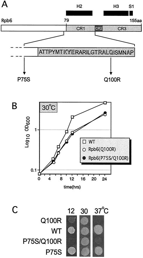
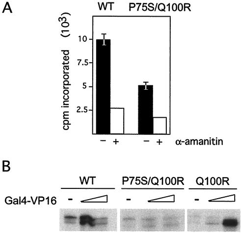
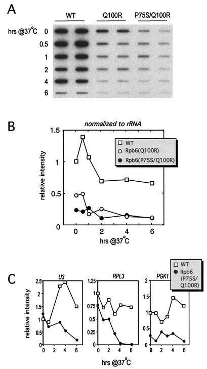
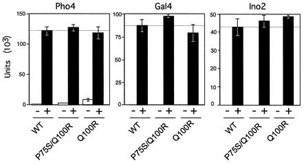
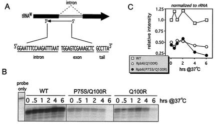
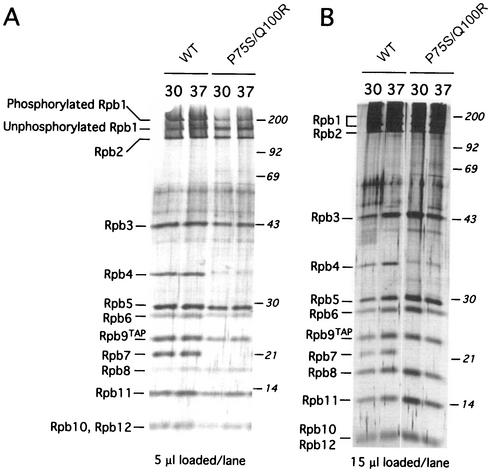
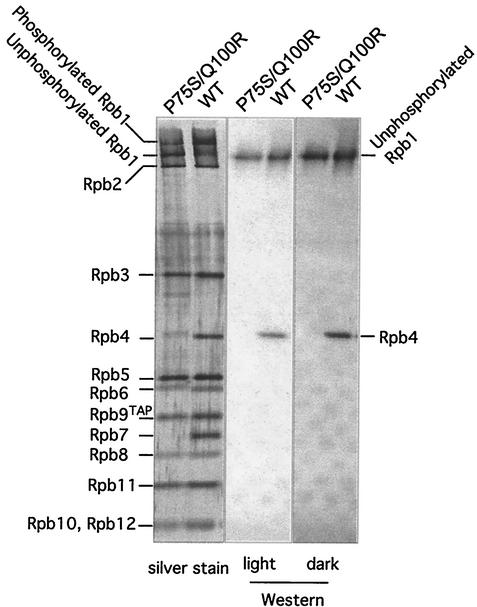
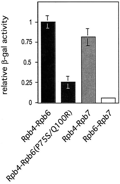
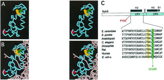
We have identified a conditional mutation in the shared Rpb6 subunit, assembled in RNA polymerases I, II, and III, that illuminated a new role that is independent of its assembly function. RNA polymerase II and III activities were significantly reduced in mutant cells before and after the shift to nonpermissive temperature. In contrast, RNA polymerase I was marginally affected. Although the Rpb6 mutant strain contained two mutations (P75S and Q100R), the majority of growth and transcription defects originated from substitution of an amino acid nearly identical in all eukaryotic counterparts as well as bacterial omega subunits (Q100R). Purification of mutant RNA polymerase II revealed that two subunits, Rpb4 and Rpb7, are selectively lost in mutant cells. Rpb4 and Rpb7 are present at substoichiometric levels, form a dissociable subcomplex, are required for RNA polymerase II activity at high temperatures, and have been implicated in the regulation of enzyme activity. Interaction experiments support a direct association between the Rpb6 and Rpb4 subunits, indicating that Rpb6 is one point of contact between the Rpb4/Rpb7 subcomplex and RNA polymerase II. The association of Rpb4/Rpb7 with Rpb6 suggests that analogous subunits of each RNA polymerase impart class-specific functions through a conserved core subunit.


Figures

FIG. 1.
FIG. 1.
Characteristics of rpb6-151. (A) Bar diagram of S. cerevisiae Rpb6. The three regions in the Rpb6 carboxy-terminal half (CR1, CR2, and CR3) displaying significant sequence similarity between all Rpb6 counterparts are delineated (gray boxes); regions of defined secondary structure (helices 2 and 3 and strand 1) are shown at top (black boxes). The complete sequence of CR1 is shown, with residues that are identical in nearly all Rpb6-related proteins in bacteria, archaebacteria, and eukaryotes highlighted (adapted from reference 32). The two amino acids mutated in rpb6-151 cells are also shown. (B) Growth curve of mutant and isogenic wild-type (WT) strains at the permissive temperature (30°C) (C) Equivalent amounts of mutant and wild-type cells were spotted onto YPD medium and incubated at the temperatures indicated.
FIG. 2.
FIG. 2.
Transcription defects in rpb6-151 cell extracts. (A) Mutant extracts have reduced activity in a promoter-independent transcription assay. Equivalent amounts of extracts were assayed for transcription on a denatured salmon sperm DNA template in the absence (−) or presence (+) of α-amanitin. Samples without α-amanitin were tested in triplicate and averaged. (B) Mutant extracts support reduced basal activity and are defective for activation in a promoter-dependent transcription assay. Shown are transcription products from wild-type (WT) and mutant whole-cell extracts with small or large amounts of Gal4-VP16 (elongated triangle) or without Gal4-VP16 (−) included in the transcription reaction. All transcription reactions used equal concentrations (300 mg) of whole-cell extracts from wild-type and mutant cells and 300 μg of template. Experiments were repeated at least three times, and the mutant phenotype was confirmed with two independently purified whole-cell extracts.
FIG. 3.
FIG. 3.
mRNA levels are significantly reduced in both Rpb6(P75S/Q100R) and Rpb6(Q100R) mutants. (A) Steady-state mRNA levels were assessed upon hybridization of radioactively labeled poly(dT)30 to 20 μg of immobilized total RNA prepared from wild-type (WT) and Rpb6 mutant cells harvested in increments after a 37°C temperature shift. (B) Band intensities in panel A were quantified and normalized to the WT 0-h time point, followed by a final normalization step to rRNA levels (as measured in Fig. 6). (C) Quantification of selected mRNA transcripts. Ten micrograms of total RNA was loaded into each lane, subjected to Northern analysis, and hybridized to the indicated DNA probes. Graphs represent quantified data from Northern analysis, with normalization to the respective 0-h time point.
FIG. 4.
FIG. 4.
Activation is not generally defective in Rpb6(P75S/Q100R) cells. Assays were performed on extracts prepared from cells harvested after growth under activating (+) or nonactivating (−) conditions. The activator whose function was tested is designated above each diagram. Values are expressed as nanomoles of ONPG cleaved per minute per milligram of protein.
FIG. 5.
FIG. 5.
Assessment of RNAP III activity. (A) tRNAw oligonucleotide sequence, components, and approximate annealing location. (B) S1 analysis of total RNA harvested at the time points indicated after a 37°C temperature shift using the probe shown in panel A. The probe-only control demonstrated that the S1 treatment was effective, since the 6-nucleotide tail was cleaved from digested samples. (C) Band intensities in panel B were quantified and normalized to the wild-type (WT) 0-h time point, followed by a final normalization step to rRNA levels (as measured in Fig. 6).
FIG. 6.
FIG. 6.
Assessment of RNAP I activity. (A) 25S rRNA oligonucleotide sequence, components, and approximate annealing location. (B) S1 analysis of total RNA harvested at the time points after a 37°C temperature shift using the probe shown in panel A. The probe-only control demonstrated that the S1 treatment was effective, since the 6-nucleotide tail was cleaved from digested samples. (C) Band intensities in panel B were quantified and normalized to the wild-type (WT) 0-h time point.
FIG. 7.
FIG. 7.
Rpb6(P75S/Q100R) is not generally defective in assembly but lacks Rpb4 and Rpb7. (A) Equivalent volumes of final purified product (amounts shown below each panel) derived from equal numbers of yeast cells harvested before and after a 2-h shift to 37°C were loaded in each lane. Molecular-weight markers are shown on the right, and positions of RNAP II subunits are shown on the left. WT, wild type. (B) Equivalent amounts of purified product were loaded in excess to clearly demonstrate the absence of the Rpb4 and Rpb7 bands.
FIG. 8.
FIG. 8.
Rpb4 is absent in Rpb6(P75S/Q100R) cells. Western analysis of mutant and wild-type (WT) samples used monoclonal antibodies to the unphosphorylated form of the CTD and to Rpb4. A silver stain of comparable samples used for Western analysis is shown on the left.
FIG. 9.
FIG. 9.
Two-hybrid analysis. Four independent experiments were performed; activities derived for interaction pairs in all four experiments were then normalized to that of the Rpb4-Rpb6 wild-type sample, averaged, and plotted as shown.
FIG. 10.
FIG. 10.
Localization of Rpb6 Q100, R97, and P75. Panels A and B (courtesy of R. Ebright, Rutgers University, Piscataway, N.J.) represent stereo pairs to enable three-dimensional viewing. (A) Structure of the conserved Rpb6 carboxy-terminal half highlighting the positions of Q100 (green), R97 (yellow), and P75 (red) displayed in Cα plus side-chain format. The structure of the amino-terminal half of Rpb6 is disordered and not included in high-resolution crystal structures derived from Protein Data Bank structure no. 1I50. (B) Rpb6 carboxy-terminal half (blue) and portions of Rpb1 (gray) represented as ribbon diagrams (also from Protein Data Bank structure no. 1I50); residues Q100, R97, and P75 are shown as in panel A. (C) Alignment of Rpb6 CR1 from an array of eukaryotes plus the Escherichia coli ω subunit. The positions of the two mutations plus R97 are highlighted in colors analogous to those used in panels A and B. Alignments are adapted from reference .

Similar articles

Cited by

References

    1. Asturias, F. J., G. D. Meredith, C. L. Poglitsch, and R. D. Kornberg. 1997. Two conformations of RNA polymerase II revealed by electron crystallography. J. Mol. Biol. 272:536-540. - PubMed
    1. Awrey, D. E., R. G. Weilbaecher, S. A. Hemming, S. M. Orlicky, C. M. Kane, and A. M. Edwards. 1997. Transcription elongation through DNA arrest sites. A multistep process involving both RNA polymerase II subunit RPB9 and TFIIS J. Biol. Chem. 272:14747-14754. - PubMed
    1. Boeke, J. D., J. Trueheart, G. Natsoulis, and G. R. Fink. 1987. 5-Fluoroorotic acid as a selective agent in yeast molecular genetics. Methods Enzymol. 154:164-175. - PubMed
    1. Burke, D., D. Dawson, and T. Stearns. 2000. Methods in yeast genetics. A Cold Spring Harbor Laboratory course manual. Cold Spring Harbor Laboratory Press, Cold Spring Harbor, N.Y.
    1. Cho, E. J., M. S. Kobor, M. Kim, J. Greenblatt, and S. Buratowski. 2001. Opposing effects of Ctk1 kinase and Fcp1 phosphatase at Ser 2 of the RNA polymerase II C-terminal domain. Genes Dev. 15:3319-3329. - PMC - PubMed

Publication types

MeSH terms