. 2003 May;23(9):3339-51.
doi: 10.1128/MCB.23.9.3339-3351.2003.

Regulation of the bone-specific osteocalcin gene by p300 requires Runx2/Cbfa1 and the vitamin D3 receptor but not p300 intrinsic histone acetyltransferase activity

Affiliations

Regulation of the bone-specific osteocalcin gene by p300 requires Runx2/Cbfa1 and the vitamin D3 receptor but not p300 intrinsic histone acetyltransferase activity

Jose Sierra et al. Mol Cell Biol. 2003 May.

Abstract

p300 is a multifunctional transcriptional coactivator that serves as an adapter for several transcription factors including nuclear steroid hormone receptors. p300 possesses an intrinsic histone acetyltransferase (HAT) activity that may be critical for promoting steroid-dependent transcriptional activation. In osteoblastic cells, transcription of the bone-specific osteocalcin (OC) gene is principally regulated by the Runx2/Cbfa1 transcription factor and is stimulated in response to vitamin D(3) via the vitamin D(3) receptor complex. Therefore, we addressed p300 control of basal and vitamin D(3)-enhanced activity of the OC promoter. We find that transient overexpression of p300 results in a significant dose-dependent increase of both basal and vitamin D(3)-stimulated OC gene activity. This stimulatory effect requires intact Runx2/Cbfa1 binding sites and the vitamin D-responsive element. In addition, by coimmunoprecipitation, we show that the endogenous Runx2/Cbfa1 and p300 proteins are components of the same complexes within osteoblastic cells under physiological concentrations. We also demonstrate by chromatin immunoprecipitation assays that p300, Runx2/Cbfa1, and 1alpha,25-dihydroxyvitamin D(3) receptor interact with the OC promoter in intact osteoblastic cells expressing this gene. The effect of p300 on the OC promoter is independent of its intrinsic HAT activity, as a HAT-deficient p300 mutant protein up-regulates expression and cooperates with P/CAF to the same extent as the wild-type p300. On the basis of these results, we propose that p300 interacts with key transcriptional regulators of the OC gene and bridges distal and proximal OC promoter sequences to facilitate responsiveness to vitamin D(3).


Figures

FIG. 1.
FIG. 1.
Schematic representation of the transcriptionally active OC promoter in an osteoblastic cell. Key regulatory elements of the rat OC gene, such as Runx2/Cbfa1 sites A, B, and C and the VDRE localized within the distal and proximal DNase I-hypersensitive sites (DHS), are shown. A nucleosome positioned between both DHS (shaded circle) and the direction of transcription (black arrow) are shown.
FIG. 2.
FIG. 2.
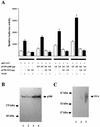
The p300 coactivator up-regulates OC gene expression. (A) ROS 17/2.8 cells were transiently cotransfected with increasing amounts of p300 expression plasmid (pCMV-p300) and 1.0 μg of 1.1-kb pOC-LUC reporter construct. The presence (+) or absence (−) and the amount of different plasmids and vitamin (Vit) D3 are shown below the bars. Cells were cultured either under control conditions (white bars) or in the presence of 10−8 M vitamin D3 (black bars) for 24 h and harvested, and luciferase activities were determined. The data were normalized to values for pCMV-β-galactosidase activity as an internal control. In some transfections, 1 μg of E1A expression vector (pCMV-E1A) was included. Pooled data from at least three independent experiments are presented. Each bar represents the mean ± standard error of the mean (n = 9; P < 0.05). (B) ROS 17/2.8 cells plated on 100-mm-diameter dishes were transiently transfected in parallel with 1.0 μg of empty vector or with increasing concentrations of the p300 expression construct. Proteins (30 μg) were resolved on SDS-8% polyacrylamide gels, transferred to nitrocellulose membranes, and the presence of p300 was revealed by Western blotting. Lane 1, 1.0 μg of empty vector; lanes 2 to 4, 0.5 μg (lane 2), 2.0 μg (lane 3), and 4.0 μg (lane 4) of p300 vector. (C) As in panel B, ROS 17/2.8 cells were transfected with empty vector or with increasing concentrations of the pE1A expression construct, and proteins were revealed by Western blotting. Lane 1, 1.0 μg of empty vector; lanes 2 and 3, 0.5 μg (lane 2) and 2.0 μg (lane 3) of E1A vector. The positions of the molecular mass markers are shown at the left of the blots.
FIG. 3.
FIG. 3.
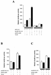
p300-dependent up-regulation of the OC promoter requires functional Runx2/Cbfa1 and VDRE sequences. Constructs (1.0 μg each) carrying wild-type (pOC-LUC), mutated Runx2/Cbfa1 sites A, B, and C (pmABC-OC-LUC) (A), mutated VDRE (pmSHE-OC-LUC) (B), or mutated Runx2/Cbfa1 sites A and B (pmAB-OC-LUC) in the context of the full-length OC promoter (1.1 kb) were cotransfected with p300 expression plasmid (3.0 μg) into ROS 17/2.8 cells. The presence (+) or absence (−) of different plasmids and vitamin (Vit) D3 are shown below the bars. Cells were cultured for 24 h in the absence (white bars) or presence (black bars) of 10−8 M vitamin D3. Luciferase reporter activities were then determined and normalized to β-galactosidase values. Pooled data from at least three independent experiments are presented. Each bar represents the mean ± standard error of the mean (n = 9; P < 0.05)
FIG. 4.
FIG. 4.
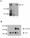
Runx2/Cbfa1 and p300 are components of the same nuclear protein complexes in osteoblastic cells expressing OC. Interaction between Runx2/Cbfa1 and p300 is shown by immunoprecipitation. Nuclear extracts from ROS 17/2.8 cells were immunoprecipitated with an anti-p300 monoclonal antibody or with a nonspecific mouse IgG fraction as described in Materials and Methods. Immunoprecipitated complexes were resolved by SDS-PAGE with 8% (A) and 10% (B) polyacrylamide gels, followed by Western blotting using either anti-p300 (A) or anti-Runx2/Cbfa1 (B) antibodies. (A) Lane 1, input; lane 2, nonspecific mouse IgG fraction; lane 3, immunoprecipitated p300. (B) Samples immunoprecipitated from ROS 17/2.8 cells cultured in the absence (lanes 1 and 3) or presence (lanes 2 and 4) of 10−8 M vitamin D3. Samples were immunoprecipitated with the nonspecific mouse IgG fraction (lanes 1 and 2) or with the p300 antibody (lanes 3 and 4). The positions of molecular mass markers are shown to the left of the blots.
FIG. 5.
FIG. 5.
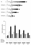
Functional interaction of Runx2/Cbfa1 and p300 requires the C-terminal region of Runx2/Cbfa1. CMV-driven constructs coding for full-length Runx2/Cbfa1 (FL) and deletion mutants of Runx2/Cbfa1 are shown schematically at the top of the figure. The Runt homology domain, the nuclear localization signal (NLS), and the nuclear matrix targeting signal (NMTS) are indicated. The functional cooperation between Runx2/Cbfa1 and p300 is demonstrated in COS-7 cells which were transiently cotransfected with different combinations of plasmids. The presence (+) or absence (−) of different plasmids is shown under the bars. The cells were transiently cotransfected with the following plasmids: CMV-empty vector (0.5 μg) (---); plasmids (0.5 μg each) coding for full-length Runx2/Cbfa1 (FL) and deletion mutant Runx2/Cbfa1 proteins (Δ432, Δ391, Δ361, and Δ230); CMV-p300 expression vector (1.0 μg); and pOC-LUC reporter construct (1.0 μg). Cells were cultured for 24 h and harvested, and luciferase activity was determined. The data were normalized to values for pCMV-β-galactosidase activity as an internal control. In some transfections, 1.0 μg of E1A expression vector (pCMV-E1A) was included.
FIG. 6.
FIG. 6.
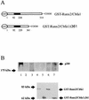
Runx2/Cbfa1 and p300 interact in vitro. The genes coding for full-length Runx2/Cbfa1 and Runx2/Cbfa1Δ361 were cloned in the pGEX-4XT1 vector and expressed as a GST fusion protein in bacteria. The abilities of these two proteins to interact with p300 in vitro were evaluated by GST pull-down experiments (see Materials and Methods). (A) Schematic illustration of the GST-Runx2/Cbfa1 and GST-Runx2/Cbfa1Δ361 proteins. The Runt homology DNA binding domain (black box) and amino acid positions are shown. (B) Glutathione-Sepharose beads (20 μl) were preincubated with 4 μg (each) of GST (lane 3), GST-Runx2/Cbfa1 (lanes 4 and 6), and GST-Runx2/Cbfa1Δ361 (lanes 5 and 7) protein for 45 min at 4°C. Nuclear extracts from ROS 17/2.8 cells (150 μg) were added to some samples (lanes 2, 3, 6, and 7) and incubated for 2 h at 4°C. The samples were then fractionated by SDS-PAGE (6% polyacrylamide gel), and the presence of bound p300 was detected by Western blot analysis. Lane 1, input; lane 2, glutathione-Sepharose plus nuclear extracts; lane 3, GST plus nuclear extracts; lane 4, GST-Runx2/Cbfa1; lane 5, GST-Runx2/Cbfa1Δ361; lane 6, GST-Runx2/Cbfa1 plus nuclear extracts; lane 7, GST-Runx2/Cbfa1Δ361 plus nuclear extracts. The bottom blot shows the results of a Western blot analysis done with an anti-Runx2/Cbfa1 polyclonal antibody to confirm that equivalent amounts of GST-Runx2/Cbfa1 and GST-Runx2/Cbfa1Δ361 are bound to the glutathione-Sepharose beads in the samples in lanes 4 to 7. The positions of the molecular mass markers are shown to the left of the blots.
FIG. 7.
FIG. 7.
p300, Runx2/Cbfa1, and VDR interact with the OC promoter in intact osteoblastic cells expressing OC. The abilities of p300, Runx2/Cbfa1, and VDR to interact with the endogenous OC promoter in intact ROS 17/2.8 cells expressing OC were determined by ChIP analysis (see Materials and Methods). (A) ChIP assays performed with formaldehyde cross-linked chromatin isolated from ROS 17/2.8 cells cultured in the absence of vitamin D3 and antibodies against p300 (left gel) and Runx2/Cbfa1 (right gel). Both gels are ethidium bromide-stained agarose gels of the PCR products obtained with OC primers with sequence from positions −459 to −28 (left gel) and positions −459 to −118 (right gel) (32) in chromatin immunoprecipitates with the indicated antibodies (p300 or Runx2). Control PCRs were done with DNA (+DNA) and without DNA (−DNA). (B) ChIP assays performed with chromatin samples from ROS 17/2.8 cells cultured in the presence (+VitD3) or absence (−VitD3) of vitamin D3 and antibodies against the VDR (left gel) or p300 (right gel). The primers for VDR (positions −773 to −433) and p300 (positions −459 to −28) were used (32). The positions of molecular size markers (in base pairs) are indicated to the left of the gels. Samples were precipitated by the nonspecific antibody IgG as a control.
FIG. 8.
FIG. 8.
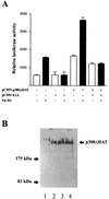
The intrinsic HAT activity of p300 is not required to up-regulate OC promoter activity. (A) An expression vector (3.0 μg) encoding a mutated p300 protein that does not display HAT activity (pCMV-p300ΔHAT) was transiently cotransfected with the pOC-LUC plasmid (1.0 μg) into ROS 17/2.8 cells cultured in the absence (white bars) or presence (black bars) of 10−8 M vitamin D3. The presence (+) or absence (−) of different plasmids and vitamin (Vit) D3 is indicated under the bars. After 24 h, the cells were harvested, and reporter activity was evaluated as described in Materials and Methods. In some transfections, the pCMV-E1A plasmid (1.0 μg) was also included. Pooled data from at least three independent experiments are presented. Each bar represents the mean ± standard error of the mean (n = 9; P < 0.05). (B) ROS 17/2.8 cells plated on 100-mm-diameter dishes were transiently transfected in parallel with either 1.0 μg of empty vector or increasing concentrations of the HAT-less p300 expression construct. Proteins (30 μg) were resolved by SDS-PAGE (8% polyacrylamide gels) and transferred to nitrocellulose membranes, and the presence of p300 was revealed by Western blotting. Lane 1, 1.0 μg of empty vector; lanes 2 to 4, 0.5 μg (lane 2), 2.0 μg (lane 3), and 4.0 μg (lane 4) of HAT-less p300 vector. The positions of molecular mass markers are shown to the left of the blot.
FIG. 9.
FIG. 9.
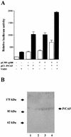
p300 cooperates with P/CAF to stimulate rat OC gene promoter activity. Expression constructs (3.0 μg each) for the coactivators p300 (wild-type p300 [A] or HAT-deficient mutant p300 [C]) and P/CAF (pCI/P/CAF) were transiently cotransfected alone or in combination in ROS 17/2.8 cells. The effect on the pOC-LUC reporter plasmid (1.0 μg) was evaluated 24 h later. Cells were cultured in the presence (black bars) or absence (white bars) of 10−8 M vitamin D3. The presence (+) and absence (−) of different plasmids and vitamin D3 (VitD3) is shown under the bars. Pooled data from at least three independent experiments are presented. Each bar represents the mean ± standard error of the mean (n = 9; P < 0.05). (B) ROS 17/2.8 cells plated on 100-mm-diameter dishes were transiently transfected in parallel with 1.0 μg of empty vector or with increasing concentrations of the P/CAF expression construct. Proteins (30 μg) were resolved by SDS-PAGE (8% polyacrylamide gels) and transferred to nitrocellulose membranes, and the presence of P/CAF was revealed by Western blotting. Lane 1, 1.0 μg of empty vector; lanes 2 to 4, 0.5 μg (lane 2), 2.0 μg (lane 3), and 4.0 μg (lane 4) of P/CAF vector. The positions of the molecular mass markers are shown to the left of the blot.
FIG. 9.
FIG. 9.
p300 cooperates with P/CAF to stimulate rat OC gene promoter activity. Expression constructs (3.0 μg each) for the coactivators p300 (wild-type p300 [A] or HAT-deficient mutant p300 [C]) and P/CAF (pCI/P/CAF) were transiently cotransfected alone or in combination in ROS 17/2.8 cells. The effect on the pOC-LUC reporter plasmid (1.0 μg) was evaluated 24 h later. Cells were cultured in the presence (black bars) or absence (white bars) of 10−8 M vitamin D3. The presence (+) and absence (−) of different plasmids and vitamin D3 (VitD3) is shown under the bars. Pooled data from at least three independent experiments are presented. Each bar represents the mean ± standard error of the mean (n = 9; P < 0.05). (B) ROS 17/2.8 cells plated on 100-mm-diameter dishes were transiently transfected in parallel with 1.0 μg of empty vector or with increasing concentrations of the P/CAF expression construct. Proteins (30 μg) were resolved by SDS-PAGE (8% polyacrylamide gels) and transferred to nitrocellulose membranes, and the presence of P/CAF was revealed by Western blotting. Lane 1, 1.0 μg of empty vector; lanes 2 to 4, 0.5 μg (lane 2), 2.0 μg (lane 3), and 4.0 μg (lane 4) of P/CAF vector. The positions of the molecular mass markers are shown to the left of the blot.

Similar articles

Cited by

References

    1. Aslam, F., L. McCabe, B. Frenkel, A. van Wijnen, G. S. Stein, J. B. Lian, and J. Stein. 1997. AP-1 and vitamin D receptor (VDR) signaling pathways converge at the rat osteocalcin VDR element: requirement for the internal activating protein-1 site for vitamin D-mediated trans-activation. Endocrinology 140:63-70. - PubMed
    1. Banerjee, C., S. Hiebert, J. Stein, J. Lian, and G. Stein. 1996. An AML-1 consensus sequence binds an osteoblast-specific complex and transcriptionally activates the osteocalcin gene. Proc. Natl. Acad. Sci. USA 93:4968-4973. - PMC - PubMed
    1. Banerjee, C., L. McCabe, J. Choi, S. Hiebert, J. Stein, G. Stein, and J. Lian. 1997. Runt homology domain proteins in osteoblast differentiation: AML3/CBFA1 is a major component of a bone-specific complex. J. Cell. Biochem. 66:1-8. - PubMed
    1. Breen, E., A. van Wijnen, J. Lian, G. Stein, and J. Stein. 1994. In vivo occupancy of the vitamin D responsive element in the osteocalcin gene supports vitamin D-dependent transcriptional upregulation in intact cells. Proc. Natl. Acad. Sci. USA 91:12902-12906. - PMC - PubMed
    1. Chakravarti, D., V. J. La Morte, M. C. Nelson, T. Nakajima, I. G. Schulman, H. Juguilon, M. Montminy, and R. M. Evans. 1996. Role of CBP/p300 in nuclear receptor signalling. Nature (London) 383:9-13. - PubMed

Publication types

MeSH terms

LinkOut - more resources