. 2003 Apr 29;100(9):5419-24.
doi: 10.1073/pnas.0830671100. Epub 2003 Apr 15.

Activation of liver X receptor improves glucose tolerance through coordinate regulation of glucose metabolism in liver and adipose tissue

Affiliations

Activation of liver X receptor improves glucose tolerance through coordinate regulation of glucose metabolism in liver and adipose tissue

Bryan A Laffitte et al. Proc Natl Acad Sci U S A. .

Abstract

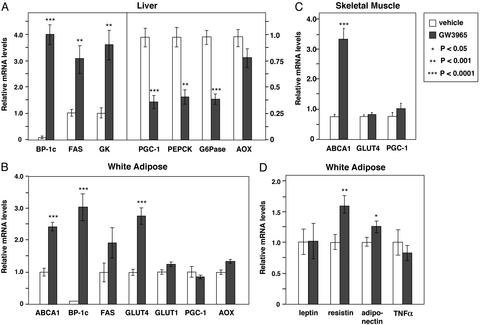
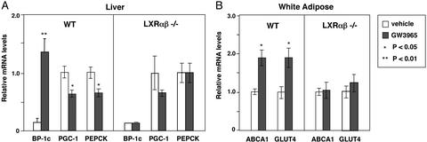
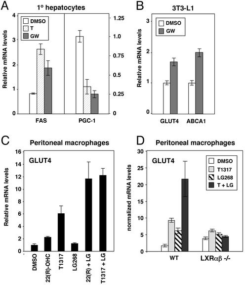
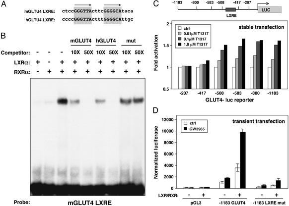
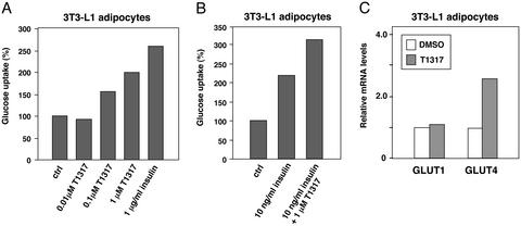
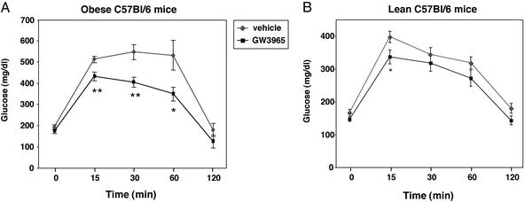
The control of lipid and glucose metabolism is closely linked. The nuclear receptors liver X receptor (LXR)alpha and LXR beta have been implicated in gene expression linked to lipid homeostasis; however, their role in glucose metabolism is not clear. We demonstrate here that the synthetic LXR agonist GW3965 improves glucose tolerance in a murine model of diet-induced obesity and insulin resistance. Analysis of gene expression in LXR agonist-treated mice reveals coordinate regulation of genes involved in glucose metabolism in liver and adipose tissue. In the liver, activation of LXR led to the suppression of the gluconeogenic program including down-regulation of peroxisome proliferator-activated receptor gamma coactivator-1 alpha (PGC-1), phosphoenolpyruvate carboxykinase (PEPCK), and glucose-6-phosphatase expression. Inhibition of gluconeogenic genes was accompanied by an induction in expression of glucokinase, which promotes hepatic glucose utilization. In adipose tissue, activation of LXR led to the transcriptional induction of the insulin-sensitive glucose transporter, GLUT4. We show that the GLUT4 promoter is a direct transcriptional target for the LXR/retinoid X receptor heterodimer and that the ability of LXR ligands to induce GLUT4 expression is abolished in LXR null cells and animals. Consistent with their effects on GLUT4 expression, LXR agonists promote glucose uptake in 3T3-L1 adipocytes in vitro. Thus, activation of LXR alters the expression of genes in liver and adipose tissue that collectively would be expected to limit hepatic glucose output and improve peripheral glucose uptake. These results outline a role for LXRs in the coordination of lipid and glucose metabolism.


Figures

Figure 1
Figure 1
Coordinate regulation of genes involved in glucose metabolism by LXR agonist in vivo. (A) LXR ligand induces glucokinase expression and represses genes involved in gluconeogenesis in liver. (B) LXR ligand induces GLUT4 expression in white adipose tissue. (C) LXR ligands regulate ABCA1 but do not alter GLUT4 or PGC-1 expression in skeletal muscle. (D) Effects of LXR ligands on expression of adipocyte signaling molecules. Ten-week-old female C57BL/6 mice (nine per group) were gavaged daily with GW3965 (20 mg/kg per day) or vehicle. At the end of the treatment period, mice were fasted for 12 h and killed, and total RNA was isolated. Gene expression for individual animals was determined by real-time quantitative-PCR assays. Results are presented as the average expression for each group ± standard deviation.
Figure 2
Figure 2
The effects of GW3965 on expression of genes involved in glucose metabolism depend on LXR expression. LXR null mice or wild-type controls on a mixed background (three to five per group) were gavaged daily with GW3965 (20 mg/kg per day) or vehicle. At the end of the treatment period, mice were fasted for 12 h and killed, and total RNA was isolated. Gene expression was determined by real-time quantitative-PCR assays. (A) Regulation of PGC-1 and PEPCK expression by GW3965 is abolished in livers of LXR null mice. (B) Regulation of GLUT4 expression is lost in adipose tissue of LXR null mice. Data are presented as expression relative to vehicle control for each genotype.
Figure 3
Figure 3
Expression of LXRs is not altered by fasting. C57BL/6 mice (four per group) were fasted for 12 h and killed, and total RNA was isolated. Gene expression was determined by real-time quantitative-PCR assays. Data are presented as relative mRNA expression.
Figure 4
Figure 4
LXR agonists regulate PGC-1 and GLUT4 expression in a cell-autonomous manner. (A) LXR ligands repress PGC-1 expression in primary human hepatocytes. (B) LXR ligand induces GLUT4 expression in 3T3-L1 adipocytes. (C) Regulation of GLUT4 expression in macrophages by synthetic and oxysterol LXR ligands. (D) Regulation of GLUT4 expression by LXR ligand is abolished in cells from LXR null mice. Cells were treated with vehicle or 1 μM T1317 (T), 1 μM GW3965 (GW), 2 μM 22(R)-hydroxycholesterol, or 50 nM LG268 for 24 h as indicated. mRNA expression was determined by real-time quantitative-PCR assays.
Figure 5
Figure 5
The GLUT4 promoter is a direct target for regulation by LXR/RXR heterodimers. (A) A conserved DR-4 hormone response element in the GLUT4 promoter. Sequence alignment of LXREs in the mouse and human GLUT4 promoters is shown. (B) A functional LXR binding site in the GLUT4 promoter. Electrophoretic mobility-shift assays were performed by using in vitro-translated proteins and radiolabeled mGLUT4 oligonucleotide. Unlabeled oligonucleotide was added as competitor as indicated. (C) LXR ligands activate the human GLUT4 promoter. Clonal populations of 3T3-L1 cells carrying stably integrated luciferase reporters under the control of various truncations of the human GLUT4 promoter were differentiated into adipocytes. On day 8 of differentiation, cells were incubated in serum-free medium with the indicated concentrations of T1317. Luciferase activity was measured 24 h later. (D) LXRE-dependent activation of the GLUT4 promoter in transient transfection assays. NIH 3T3 cells were transfected with wild-type or LXRE mutant −1,183-bp human GLUT4 luciferase reporters along with expression vectors for LXRα and RXRα. Cells were treated with 1 μM GW3965 12 h after transfection, and luciferase activity was determined 24 h later.
Figure 6
Figure 6
Synthetic LXR ligand promotes glucose uptake in 3T3-L1 adipocytes. (A and B) 3T3-L1 adipocytes at day 10 of differentiation were incubated for 24 h with serum-free medium supplemented with T1317 and/or insulin as indicated. The following day, glucose uptake of treated cells was measured in 96-well Cytostar-T microplates using 14C-labeled 2-deoxyglucose. (C) LXR agonist increases GLUT4 mRNA expression in 3T3-L1 cells. Gene expression was determined by real-time quantitative-PCR assays.
Figure 7
Figure 7
Synthetic LXR ligand improves glucose tolerance in a model of diet-induced obesity and insulin resistance. (A) Effect of GW3965 on glucose tolerance in obese mice. C57BL/6 mice were fed a high-fat diet for 3 months to induce obesity. After 3 months, mice were treated for 1 week with vehicle or 20 mg/kg body weight GW3965. Glucose-tolerance tests were performed by i.p. injection of glucose (2 g/kg body weight) after 8 h of fasting. (B) Effect of GW3965 on glucose tolerance in lean C57BL/6 mice. Lean C57BL/6 mice were maintained on normal chow diet and treated as described above. *, P < 0.05; **, P < 0.01.

Similar articles

Cited by

References

    1. Janowski B A, Willy P J, Devi T R, Falck J R, Mangelsdorf D J. Nature. 1996;383:728–731. - PubMed
    1. Lehmann J M, Kliewer S A, Moore L B, Smith-Oliver T A, Oliver B B, Su J L, Sundseth S S, Winegar D A, Blanchard D E, Spencer T A, Willson T M. J Biol Chem. 1997;272:3137–3140. - PubMed
    1. Fu X, Menke J G, Chen Y, Zhou G, MacNaul K L, Wright S D, Sparrow C P, Lund E G. J Biol Chem. 2001;276:38378–38387. - PubMed
    1. Repa J J, Mangelsdorf D J. Annu Rev Cell Dev Biol. 2000;16:459–481. - PubMed
    1. Repa J J, Mangelsdorf D J. Nat Med. 2002;8:1243–1248. - PubMed

Publication types

MeSH terms

LinkOut - more resources