. 2003 Apr 15;28(8):755-63.

Potential use of Sox9 gene therapy for intervertebral degenerative disc disease

Affiliations

Potential use of Sox9 gene therapy for intervertebral degenerative disc disease

Ronjon Paul et al. Spine (Phila Pa 1976). .

Abstract

Study design: A new recombinant adenoviral vector expressing Sox9, a chondrocyte-specific transcription factor, was tested in a chondroblastic cell line and primary human intervertebral disc cells in vitro. Direct infection of intervertebral disc cells then was assessed in a rabbit model.

Objectives: To deliver a potentially therapeutic viral vector expressing Sox9 to degenerative human and rabbit intervertebral discs cells, and to assess the effect of Sox9 expression on Type 2 collagen production.

Summary of the background data: The concentration of competent Type 2 collagen, an essential constituent of the healthy nucleus pulposus, declines with intervertebral disc degeneration. Recent studies suggest that Sox9 upregulates Type 2 collagen production. Interventions that augment Type 2 collagen production by intervertebral disc cells may represent a novel therapeutic method for patients with degenerative disc disease.

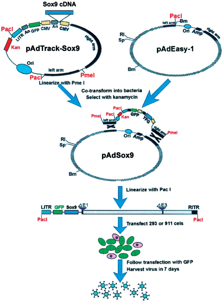
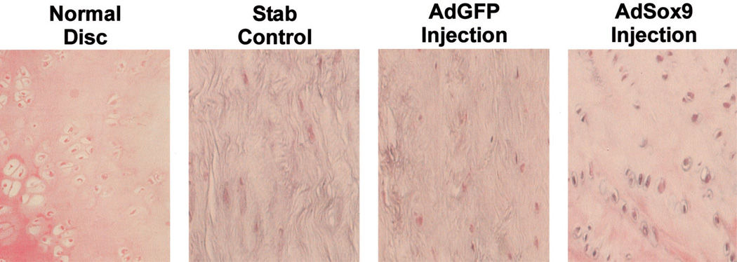
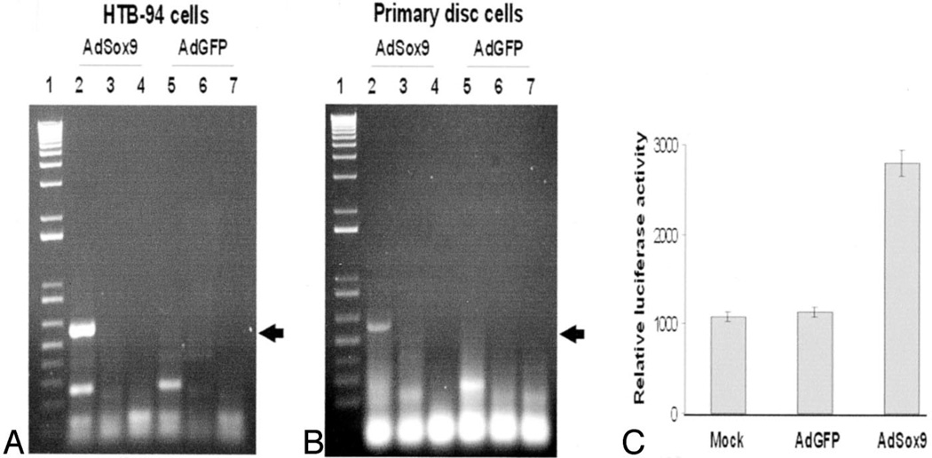
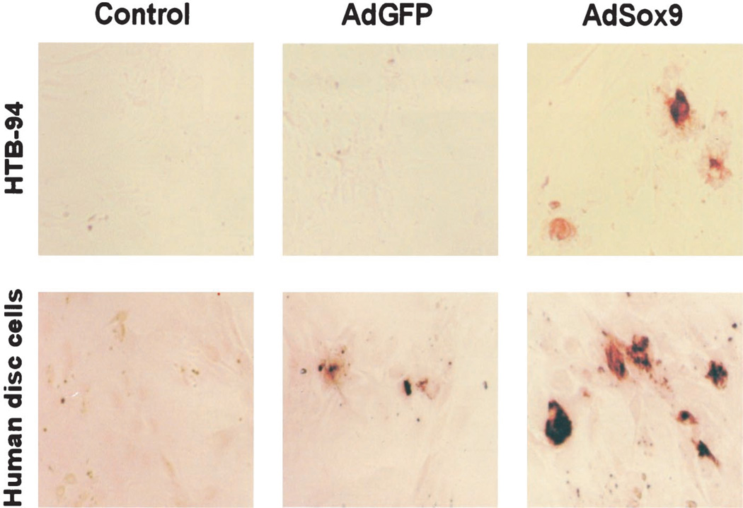
Methods: Adenoviral delivery vectors expressing Sox9 and green fluorescent protein were constructed using the AdEasy system. The chondroblastic cell line, HTB-94, and cultured human degenerated intervertebral disc cells were infected with the vectors. Reverse transcriptase-polymerase chain reaction and immunohistochemical analyses were performed to document increased Type 2 collagen expression. The AdSox9 virus then was injected directly into the intervertebral discs of three rabbits. After 5 weeks, the injected discs were evaluated histologically.

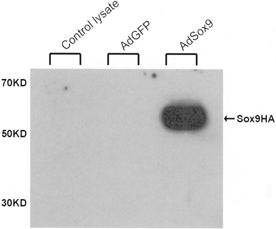
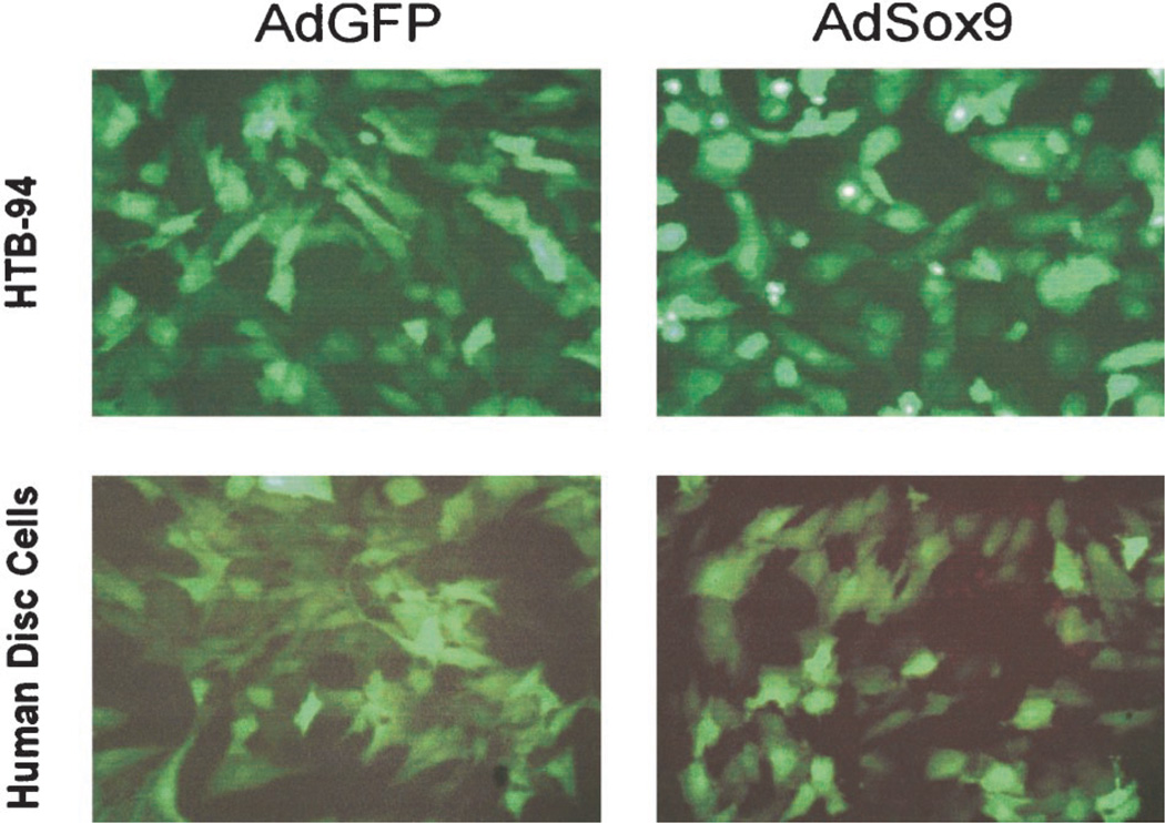
Results: The AdSox9 virus efficiently transduced HTB-94 cells and degenerated human disc cells. Western blot analysis confirmed increased Sox9 production. Increased Type 2 collagen production was demonstrated in infected HTB-94 and human disc cells using both reverse transcriptase-polymerase chain reaction and immunohistochemical staining. In the rabbit model, cells infected with AdSox9 maintained a chondrocytic phenotype, and the architecture of the nucleus pulposus was preserved over a 5-week study period compared to control discs.

Conclusions: A novel adenoviral vector efficiently increased Sox9 and Type 2 collagen synthesis in cultured chondroblastic cells and human degenerated disc cells. In a rabbit model, sustained Sox9 production preserved the histologic appearance of the nucleus pulposus cells in vivo. These findings suggest a potential role for Sox9 gene therapy in the treatment of human degenerative disc disease.


Conflict of interest statement

Conflict of interest: No funds were received in support of this work. No benefits in any form have been or will be received from a commercial party related directly or indirectly to the subject of this article.

Figures

Figure 1
Figure 1
Schematic representation of adenoviral vector AdSox9 construction. An HA-tagged cDNA coding sequence of human Sox9 was subcloned into a shuttle vector, pAdTracK-CMV. Resultant pAdTrack-Sox9 was next used to generate adenoviral recombinants through homologous recombination with the ad-enoviral backbone vector, pAdEasy-1, in BJ5183 bacterial cells. After being linearized with Pac I, the adenoviral recombi-nants were used to produce ad-enoviruses in HEK 293 packaging cells, resulting in an AdSox9 ad-enoviral vector that contained a built-in GFP expression cassette. A recombinant adenoviral vector expressing GFP alone also was constructed as a control vector (AdGFP). For detailed information about AdEasy System, please refer to He et al., Proc Natl Acad Sci (AM) 1998;95:2509, and www.coloncancer.org/adeasy.htm
Figure 2
Figure 2
Intervertebral disc degeneration induced by needle anulotomy in a rabbit model. A 27-gauge needle puncture through the anterior anulus into the nucleus pulposus was performed. Certain discs then were injected with 10 µL of saline. At 6 weeks after injection, the animals were killed and the lumbar segments retrieved. Macrographic images were prepared from the intact and hemisected specimens. At anulotomy and anulotomy followed by saline-injection of discs, disc space narrowing and osteophyte formation is evident at 6 weeks. Effect of Sox9 on intervertebral disc tissue in rabbits. Recombinant adenoviruses (1 × 10⁹ pfu) were injected into injured rabbit lumbar intervertebral disc. Three test conditions were studied: 1) control (stab incision without injection), 2) AdGFP (stab incision with injection of mock adenovirus, AdGFP), and 3) AdSox9 (stab incision with injection of AdSox9 virus). At 5 weeks after injection, the animals were killed. The lumbar disc tissues were recovered and stained with hematoxylin–eosin. A normal, uninjured disc was included for comparison. The disc that underwent annulotomy without injection of adenovirus as well as the disc that underwent annulotomy with injection of the mock virus AdGFP, demonstrated replacement of the disc with fibrocytic cells and dense extracellular matrix. The disc that received the AdSox9 virus retained the histologic appearance of cartilage.
Figure 3
Figure 3
Expression of exogenous Sox9 mediated by the AdSox9 adenoviral vector. Subconfluent HCT116 cells were infected with AdSox9 or AdGFP. Total cell lysate was collected 24 hours after infection and subjected to 4% to 20% SDS–SPAGE (approximately 10 µg total proteins per lane). After being resolved and transferred to an Immobilon-P membrane, the presence of HA-tagged Sox9 protein was probed with a mouse monoclonal anti-HA antibody (12CA5, Roche Molecular Biochemicals) and visualized using the SuperSignal West Pico chemiluminescent substrate kit (PIERCE).
Figure 4
Figure 4
Efficient transduction of chondrocytes by recombinant adenoviral vectors. Subconfluent HTB-94 and primary human disc cells were infected with AdSox9 or AdGFP. At 24 hours of adenoviral infection, expression of GFP was visualized and recorded using fluorescence microscopy.
Figure 5
Figure 5
Induction of Type 2 collagen expression by exogenous Sox9. A and B, RT-PCR analysis of Type 2 collagen expression. Exponentially growing HTB-94 cells (A) and primary disc cells (B) were infected with AdSox9 and AdGFP. At 48 hours after infection, total RNA was isolated and subjected to reverse transcriptase-PCR reactions. Expression of Type 2 collagen was detected by PR-PCR analysis using a pair of Type 2 collagen-specific primers. The expected PCR product was approximately 560 bp (arrows). Lane 1: 1-kb plus ladder (Life Technologies). Lanes 2 and 5: templates derived from +RT reactions. Lanes 3 and 6: no template control. Lanes 4 and 7: templates derived from −RT reactions. C, Activation of Type 2 collagen promoter-containing reporter. Subconfluent HTB-94 cells were transfected with a reporter construct, pGL2–3.774kb, that contained a luciferase reporter gene driven by human COL2A1 promoter. At 20 hours after transfection, the cells were replated and infected with AdSox9, AdGFP, or no infection (mock). At 48 hours after infection, the cells were lysed and collected for luciferase activity assays using the Luciferase Assay kit (Promega). Each assay condition was performed in triplicate.
Figure 6
Figure 6
Immunohistochemical analysis of Type 2 collagen production induced by exogenous Sox9. Subconfluent HTB-94 and primary disc cells were infected with AdSox9 or AdGFP. At 36 hours after infection, the cells were fixed and immunostained with a rat anti-Type 2 collagen antibody (provided by Dr. Michael Cremer, University of Tennessee, Memphis, TN). The control condition represented infected cells immunostained without the primary antibody.

References

    1. Borenstein D. Epidemiology, etiology, diagnostic evaluation, and treatment of low back pain. Curr Opin Rheumatol. 1992;4:226–232. - PubMed
    1. Frymoyer JW, Cats-Baril WL. An overview of the incidences and costs of low back pain. Orthop Clin North Am. 1991;22:263–271. - PubMed
    1. Webster BS, Snook SH. The cost of 1989 workers’ compensation low back pain claims. Spine. 1994;19:1111–1115. discussion 1116. - PubMed
    1. Antoniou J, Steffen T, Nelson F, et al. The human lumbar intervertebral disc: Evidence for changes in the biosynthesis and denaturation of the extracellular matrix with growth, maturation, ageing, and degeneration. J Clin Invest. 1996;98:996–1003. - PMC - PubMed
    1. Buckwalter JA. Aging and degeneration of the human intervertebral disc. Spine. 1995;20:1307–1314. - PubMed

Publication types

MeSH terms