Biased T-cell receptor repertoires in patients with chromosome 22q11.2 deletion syndrome (DiGeorge syndrome/velocardiofacial syndrome)
- PMID: 12699424
- PMCID: PMC1808695
- DOI: 10.1046/j.1365-2249.2003.02134.x
Biased T-cell receptor repertoires in patients with chromosome 22q11.2 deletion syndrome (DiGeorge syndrome/velocardiofacial syndrome)
Abstract
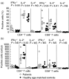
Chromosome 22q11.2 deletion (del22q11.2) syndrome (DiGeorge syndrome/velocardiofacial syndrome) is a common syndrome typically consisting of congenital heart disease, hypoparathyroidism, developmental delay and immunodeficiency. Although a broad range of immunologic defects have been described in these patients, limited information is currently available on the diversity of the T-cell receptor (TCR) variable beta (BV) chain repertoire. The TCRBV repertoires of nine patients with del22q11.2 syndrome were determined by flow cytometry, fragment size analysis of the third complementarity determining region (CDR3 spectratyping) and sequencing of V(D)J regions. The rate of thymic output and the phenotype and function of peripheral T cells were also studied. Expanded TCRBV families were detected by flow cytometry in both CD4+ and CD8+ T cells. A decreased diversity of TCR repertoires was also demonstrated by CDR3 spectratyping, showing altered CDR3 profiles in the majority of TCRBV families investigated. The oligoclonal nature of abnormal peaks detected by CDR3 spectratyping was confirmed by the sequence analysis of the V(D)J regions. Thymic output, evaluated by measuring TCR rearrangement excision circles (TRECs), was significantly decreased in comparison with age-matched controls. Finally, a significant up-regulation in the percentage, but not in the absolute count, of activated CD4+ T cells (CD95+, CCR5+, HLA-DR+), IFN-gamma - and IL-2-expressing T cells was detected. These findings suggest that the diversity of CD4 and CD8 TCRBV repertoires is decreased in patients with del22q11.2 syndrome, possibly as a result of either impaired thymic function and/or increased T-cell activation.
Figures


Similar articles
-
Post-natal ontogenesis of the T-cell receptor CD4 and CD8 Vbeta repertoire and immune function in children with DiGeorge syndrome.J Clin Immunol. 2005 May;25(3):265-74. doi: 10.1007/s10875-005-4085-3. J Clin Immunol. 2005. PMID: 15981092
-
Flow cytometric analysis of TCR Vβ repertoire in patients with 22q11.2 deletion syndrome.Scand J Immunol. 2011 Jun;73(6):577-85. doi: 10.1111/j.1365-3083.2011.02527.x. Scand J Immunol. 2011. PMID: 21323691
-
T-cell homeostasis in humans with thymic hypoplasia due to chromosome 22q11.2 deletion syndrome.Blood. 2004 Feb 1;103(3):1020-5. doi: 10.1182/blood-2003-08-2824. Epub 2003 Oct 2. Blood. 2004. PMID: 14525774
-
Thymic commitment of regulatory T cells is a pathway of TCR-dependent selection that isolates repertoires undergoing positive or negative selection.Curr Top Microbiol Immunol. 2005;293:43-71. doi: 10.1007/3-540-27702-1_3. Curr Top Microbiol Immunol. 2005. PMID: 15981475 Review.
-
Human T cell reconstitution in DiGeorge syndrome and HIV-1 infection.Semin Immunol. 2007 Oct;19(5):297-309. doi: 10.1016/j.smim.2007.10.002. Epub 2007 Nov 26. Semin Immunol. 2007. PMID: 18035553 Free PMC article. Review.
Cited by
-
Neonatal Levels of T-cell Receptor Excision Circles (TREC) in Patients with 22q11.2 Deletion Syndrome and Later Disease Features.J Clin Immunol. 2015 May;35(4):408-15. doi: 10.1007/s10875-015-0153-5. Epub 2015 Mar 27. J Clin Immunol. 2015. PMID: 25814142
-
Primary and secondary defects of the thymus.Immunol Rev. 2024 Mar;322(1):178-211. doi: 10.1111/imr.13306. Epub 2024 Jan 16. Immunol Rev. 2024. PMID: 38228406 Free PMC article. Review.
-
Secondary immunologic consequences in chromosome 22q11.2 deletion syndrome (DiGeorge syndrome/velocardiofacial syndrome).Clin Immunol. 2010 Sep;136(3):409-18. doi: 10.1016/j.clim.2010.04.011. Epub 2010 May 15. Clin Immunol. 2010. PMID: 20472505 Free PMC article.
-
The TREC/KREC assay for the diagnosis and monitoring of patients with DiGeorge syndrome.PLoS One. 2014 Dec 8;9(12):e114514. doi: 10.1371/journal.pone.0114514. eCollection 2014. PLoS One. 2014. PMID: 25485546 Free PMC article.
-
COVID-19 Severity, Cardiological Outcome, and Immunogenicity of mRNA Vaccine on Adult Patients With 22q11.2 DS.J Allergy Clin Immunol Pract. 2023 Jan;11(1):292-305.e2. doi: 10.1016/j.jaip.2022.10.010. Epub 2022 Oct 21. J Allergy Clin Immunol Pract. 2023. PMID: 36280136 Free PMC article.
References
-
- Edelmann L, Pandita RK, Spiteri E, et al. A common molecular basis for rearrangement disorders on chromosome 22q11. Hum Mol Genet. 1999;8:1157–67. - PubMed
-
- Yamagishi H, Ishii C, Maeda J, et al. Phenotypic discordance in monozygotic twins with 22q11.2 deletion. Am J Med Genet. 1998;78:319–21. - PubMed
-
- Novelli G, Amati F, Dallapiccola B. Individual haploinsufficient loci and the complex phenotype of DiGeorge syndrome. Mol Med Today. 2000;6:10–1. - PubMed
Publication types
MeSH terms
Substances
Grants and funding
LinkOut - more resources
Full Text Sources
Research Materials

