. 2003 May 1;12(9):961-73.
doi: 10.1093/hmg/ddg119.

Production of MPS VII mouse (Gus(tm(hE540A x mE536A)Sly)) doubly tolerant to human and mouse beta-glucuronidase

Affiliations

Production of MPS VII mouse (Gus(tm(hE540A x mE536A)Sly)) doubly tolerant to human and mouse beta-glucuronidase

Shunji Tomatsu et al. Hum Mol Genet. .

Abstract

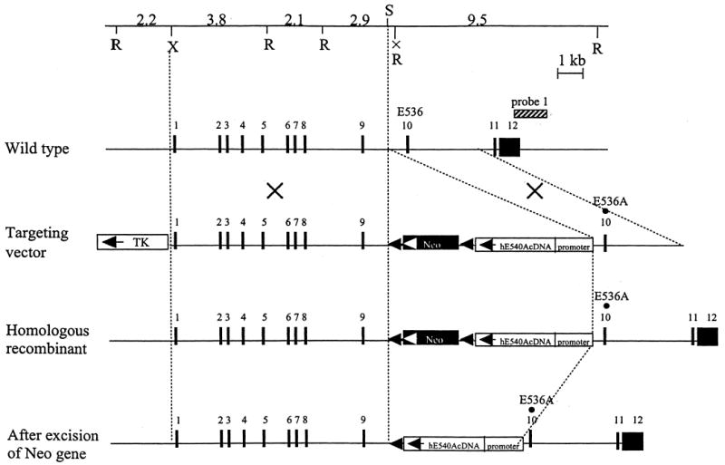
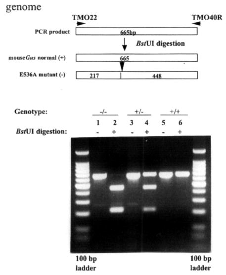
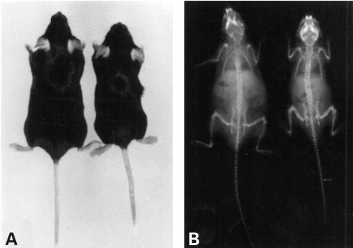
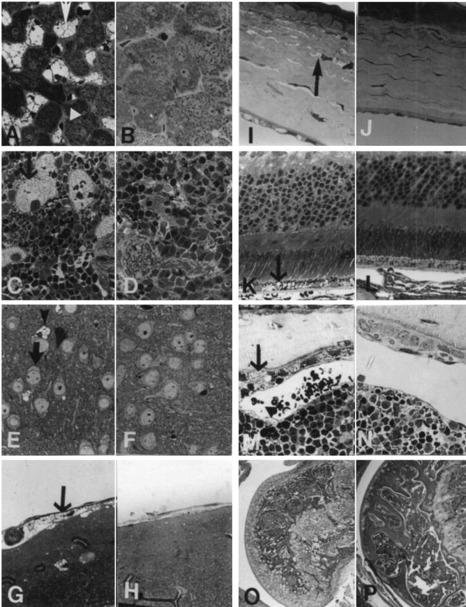
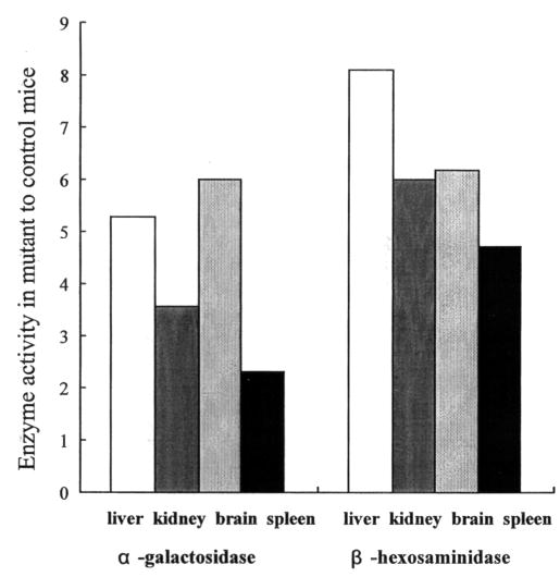
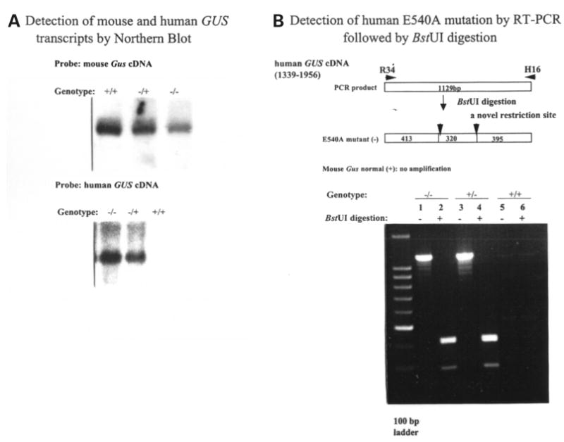
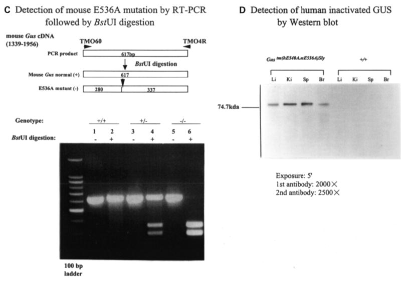
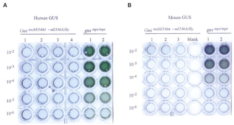
Mucopolysaccharidosis VII (MPS VII, Sly syndrome) is an autosomal recessive lysosomal storage disease caused by beta-glucuronidase (GUS) deficiency. A naturally occurring mouse model of that disease has been very useful for studying experimental approaches to therapy. However, immune responses can complicate evaluation of the long-term benefits of enzyme replacement or gene therapy delivered to adult MPS VII mice. To make this model useful for studying the long-term effectiveness and side effects of experimental therapies delivered to adult mice, we developed a new MPS VII mouse model, which is tolerant to both human and murine GUS. To achieve this, we used homologous recombination to introduce simultaneously a human cDNA transgene expressing inactive human GUS into intron 9 of the murine Gus gene and a targeted active site mutation (E536A) into the adjacent exon 10. When the heterozygote products of germline transmission were bred to homozygosity, the homozygous mice expressed no GUS enzyme activity but expressed inactive human GUS protein highly and were tolerant to immune challenge with human enzyme. Expression of the mutant murine Gus gene was reduced to about 10% of normal levels, but the inactive murine GUS enzyme also conferred tolerance to murine GUS. This MPS VII mouse model should be useful to evaluate therapeutic responses in adult mice receiving repetitive doses of enzyme or mice receiving gene therapy as adults. Heterozygotes expressed only 9.5-26% of wild-type levels of murine GUS instead of the expected 50%, indicating a dominant-negative effect of the mutant enzyme monomers on the activity of GUS tetramers in different tissues. Corrective gene therapy in this model should provide high enough levels of expression of normal GUS monomers to overcome the dominant negative effect of mutant monomers on newly synthesized GUS tetramers in most tissues.


Figures

Figure 1
Figure 1
Targeted mutagenesis of the Gus gene. The structure of the endogenous gene, the targeting construct, the homologous recombinant allele, and the neo-excised allele are presented schematically on successive lines. Filled rectangles represent exons and neor, whereas two open rectangles indicate TK and human GUS cDNA, respectively. The striped bar over the wild-type allele represents the probe used for Southern blots. Abbreviations for restriction enzymes are R, EcoRI; S, SalI; X, XhoI. The EcoRI site in intron 9 (X) was lost during the construction of the targeting vector by in vitro mutagenesis without any effect on the consensus splicing sequences. The homologous E to A amino acid change was introduced in both the mouse Gus gene (E536) and the human GUS cDNA (E540).
Figure 2
Figure 2
Detection of E536A point mutation in the murine Gus gene by genomic PCR amplification and subsequent BstUI digestion. The E536A mutation (an A→C transversion) creates a new restriction site, BstUI. Restriction enzyme analysis of the mutation introduced at codon 536, E536A, was performed using DNA from Gustm(hE540A·mE536A)Sly (−/−), Gustm(hE540A·mE536A)Sly/+ (+/−), and Gus+/+ (+/+) mice. Unnumbered lanes at each end are DNA ladders of 100 bp markers. Lane 1, undigested amplified PCR product (665 bp) from an E536A homozygote (−/−); lane 2, DNA from an E536A homozygote digested with BstUI (217 and 448 bp); lane 3, undigested amplified PCR product from a heterozygote (+/−); lane 4, DNA from a heterozygote digested with BstUI (217, 448 and 665 bp); lane 5, undigested amplified PCR product from a wild-type control (+/+); lane 6, DNA from a wild-type control digested with BstUI.
Figure 3
Figure 3
Phenotype of MPS VII doubly tolerant mouse. (A) A 4-month-old MPS VII doubly tolerant female mouse (right) compared with a 4-month-old normal female (left). The MPS VII mouse is smaller than the normal mouse and has shortened limbs, a hobbled gait, and a dysmorphic face with a blunted nose. (B) The skeleton of a 4-month-old MPS VII doubly tolerant male mouse (right) shows sclerosis of the cranial bones, a broad zygomatic arch, shortened limb bones, and a narrow rib cage, compared with the skeleton of a normal 4-month-old male mouse (left).
Figure 4
Figure 4
Morphological alteration in the MPS VII doubly tolerant Gustm(hE540 A·mE536A)Sly mice. (A) Liver from an 8-month-old Gustm(hE540A·mE536A)Sly mouse has Kupffer cells that are distended with lysosomal storage (arrow). The hepatocytes have only a small amount of cytoplasmic storage (arrowhead). (B) Neither hepatocytes nor Kupffer cells were altered in an adult heterozygote Gustm(hE540A.mE536A)Sly/+ mouse. (C) Spleen from a 7-month-old Gustm(hE540A·mE536A)Sly mouse has prominent lysosomal storage in the sinus lining cells. (D) No storage is apparent in the spleen of a Gustm(hE540A·mE536A)Sly/+ mouse. (E) The neocortical neurons (arrow) and glial cells (arrowhead) in a 1-month-old Gustm(hE540A.mE536A)Sly mouse have lysosomal distention. (F) The neocortex in a Gustm(hE540A·mE536A)Sly/+ mouse has no evidence of lysosomal storage in either neurons or in glial cells. (G) The meninges covering the brain contain cells distended with lysosomal storage (arrow) in a 1-month-old Gustm(hE540A·mE536A)Sly mouse. (H) In the meninges of a Gustm(hE540A·mE536A)Sly/+ mouse there is no evidence of lysosomal storage. (I) The cornea from a 1-month-old Gustm(hE540A·mE536A)Sly mouse has stromal fibrocytes (arrow) with a moderate amount of lysosomal distention. (J) The Gustm(hE540A·mE536A)Sly/+ mouse has no storage in the corneal fibrocytes or epithelium. (K) The retinal pigment epithelium at the base of the retina in a 1-month-old Gustm(hE540A·mE536A)Sly mouse is distended with storage (arrow). Other layers of the retina have no lysosomal storage accumulation apparent at the light microscopic level. (L) The retina from a Gustm(hE540A·mE536A)Sly/+ mouse has no morphological abnormality. (M) Bone from the rib of a 2-month-old Gustm(hE540A·mE536A)Sly mouse shows distended osteoblasts lining the cortical bone (arrow) and osteocytes within the bone with a moderate amount of lysosomal distention. The sinus lining cells in bone marrow (arrowhead) also contain a small amount of storage. (N) Neither the bone marrow nor the bone had a morphological alteration in the Gustm(hE540A·mE536A)Sly/+ mice. (O) A stifle joint from the limb of an 8-month-old Gustm(hE540A·mE536A)Sly mouse shows the distortion of the bone architecture. There is storage and structural alteration of both the articular and epiphyseal cartilage plate chondrocytes. (P) A similar joint from an adult Gustm(hE540A·mE536A)Sly/+ mouse has no structural alteration. (A–N, toluidine blue, 1 cm=27 μm; O,P hematoxylin and eosin, 1 cm=425 μm).
Figure 5
Figure 5
Storage in cerebellum of control and MPS mice. (A) A cerebellar Purkinje cell in a wild-type control mouse has normal cytoplasm with no evidence of storage. (B) A Purkinje cell from a homozygous mouse contains several large membrane-bound accumulations of flocculent fibrillar material, identical to the storage material seen in the previously described Birkenmeier MPS VII murine model. (C) A Purkinje cell from a heterozygous mouse contains membrane-bound stored material that is similar, although slightly more complex ultrastructurally, than that seen in the homozygous mutant mouse (A, B and C, uranyl acetate–lead citrate; 2080×).
Figure 6
Figure 6
Secondary elevation of α-galactosidase and β-hexosaminidase. Levels of α-galactosidase and β-hexosaminidase in tissues of Gustm(hE540A·mE536A)Sly mice, expressed as fold increase over levels found in B6 control mice. Normal B6 control mean α-galactosidase levels in liver, kidney, brain and spleen are 45, 28, 19 and 67 units/μg protein, respectively. Normal B6 control mean β-hexosaminidase levels in liver, kidney, brain and spleen are 442, 781, 876 and 2525 units/μg protein, respectively.
Figure 7
Figure 7
Expression of murine Gus (or human GUS) mRNA and GUS protein. (A) Northern blot analysis of murine Gus (upper panel) or human GUS (lower panel) mRNA from the livers of wild-type (+/+), heterozygote (+/−) and Gustm(hE540A·mE536A)Sly homozygote (−/−) mice. The 2.3 kb murine Gus mRNA transcript present in homozygous Gustm(hE536A·mE536A)Sly liver (upper panel, lane 3) was reduced in comparison to wild-type (+/+) and heterozygote (+/−) mice (upper panel, lanes 1 and 2). The murine probe does not cross hybridize with the human transcript under these conditions (data not shown). The human GUS mRNA was expressed only in the transgene containing Gustm(hE540A·mE536A)Sly homozygote and heterozygote tissues (lower panel, lanes 1 and 2). (B) BstUI analysis of human E540A mutation in hGUS transgene using RT–PCR. Fragments of 1129 bp were amplified from Gustm(hE540A·mE536A)Sly and heterozygote mice (lanes 1 and 3) using primers R34 and H16 (and visualized on a 2% agarose gel). No amplification from the wild-type mouse DNA (lanes 5 and 6) were seen. The 1129 bp fragments amplified from the mutant human transgene were cleaved into 413, 320 and 395 bp fragments by digestion (lanes 2 and 4). Note that the 413 and 395 bp fragments do not separate, so a doublet of these two and a single 320 bp band are seen. (C) BstUI analysis of mouse genomic DNA for the E536A mutation using RT–PCR. The 617 bp fragments were amplified using primers TMO60 and TMO4R from the homozygous Gustm(hE540A·mE536A)Sly (lanes 5 and 6), wild-type (lanes 1 and 2), and heterozygous Gustm(hE540A·mE536A)Sly/+ (lanes 3 and 4) mice, respectively. BstUI digestion cleaves the products from the mutant allele into 280 and 337 bp fragments (lanes 4 and 6). The 617 bp product of the normal allele is not cleaved by BstUI (lane 2). (D) Western blot for human GUS protein in extracts of tissues from Gustm(hE540A·mE536A)Sly and wild-type mice. The hGUS proteins from liver, kidney, spleen and brain tissues of the Gustm(hE540A·mE536A)Sly mouse were identified on western blot by anti-human GUS antibody (lanes 1–4). No band was observed in any tissue from the control wild-type mouse.
Figure 7
Figure 7
Expression of murine Gus (or human GUS) mRNA and GUS protein. (A) Northern blot analysis of murine Gus (upper panel) or human GUS (lower panel) mRNA from the livers of wild-type (+/+), heterozygote (+/−) and Gustm(hE540A·mE536A)Sly homozygote (−/−) mice. The 2.3 kb murine Gus mRNA transcript present in homozygous Gustm(hE536A·mE536A)Sly liver (upper panel, lane 3) was reduced in comparison to wild-type (+/+) and heterozygote (+/−) mice (upper panel, lanes 1 and 2). The murine probe does not cross hybridize with the human transcript under these conditions (data not shown). The human GUS mRNA was expressed only in the transgene containing Gustm(hE540A·mE536A)Sly homozygote and heterozygote tissues (lower panel, lanes 1 and 2). (B) BstUI analysis of human E540A mutation in hGUS transgene using RT–PCR. Fragments of 1129 bp were amplified from Gustm(hE540A·mE536A)Sly and heterozygote mice (lanes 1 and 3) using primers R34 and H16 (and visualized on a 2% agarose gel). No amplification from the wild-type mouse DNA (lanes 5 and 6) were seen. The 1129 bp fragments amplified from the mutant human transgene were cleaved into 413, 320 and 395 bp fragments by digestion (lanes 2 and 4). Note that the 413 and 395 bp fragments do not separate, so a doublet of these two and a single 320 bp band are seen. (C) BstUI analysis of mouse genomic DNA for the E536A mutation using RT–PCR. The 617 bp fragments were amplified using primers TMO60 and TMO4R from the homozygous Gustm(hE540A·mE536A)Sly (lanes 5 and 6), wild-type (lanes 1 and 2), and heterozygous Gustm(hE540A·mE536A)Sly/+ (lanes 3 and 4) mice, respectively. BstUI digestion cleaves the products from the mutant allele into 280 and 337 bp fragments (lanes 4 and 6). The 617 bp product of the normal allele is not cleaved by BstUI (lane 2). (D) Western blot for human GUS protein in extracts of tissues from Gustm(hE540A·mE536A)Sly and wild-type mice. The hGUS proteins from liver, kidney, spleen and brain tissues of the Gustm(hE540A·mE536A)Sly mouse were identified on western blot by anti-human GUS antibody (lanes 1–4). No band was observed in any tissue from the control wild-type mouse.
Figure 8
Figure 8
Humoral immune tolerance of Gustm(hE540A·mE536A)Sly mice to human and murine GUSs. (A) ELISA plate assay of antibodies to hGUS in serum of Gustm(hE540A·mE536A)Sly mice (left, lanes 1–4) and control MPS VII (gusmps/mps) mice (right, lanes 1 and 2) following primary immunization with human GUS in complete Freund’s adjuvant and two boosts with human GUS in incomplete Freund’s adjuvant. Gustm(hE540A·mE536A)Sly mice show no antibody response whereas control mice have antibodies detectable at 105 dilutions or greater. (B) ELISA plate assay of antibodies to mGUS in serum of Gustm(hE540A·mE536A)Sly mice (left, lanes 1–3) and control MPS VII (gusmps/mps) mice (right, lanes 1 and 2) following primary immunization with mouse GUS in complete Freund’s adjuvant and two boosts with mouse GUS in incomplete Freund’s adjuvant.

Similar articles

Cited by

References

    1. Sly WS, Quinton BA, McAlister WH, Rimoin DL. Beta-glucuronidase deficiency: report of clinical, radiologic, and biochemical features of a new mucopolysaccharidosis. J Pediatr. 1973;82:249–257. - PubMed
    1. Neufeld EF, Muenzer J. The mucopolysaccharidoses. In: Scriver CR, Beaudet AL, Sly WS, Valle D, editors. The Metabolic and Molecular Bases of Inherited Disease. Vol. 3. McGraw-Hill, Medical Publishing Division; New York: 2001. pp. 3421–3452.
    1. Paigen K. Mammalian beta-glucuronidase: genetics, molecular biology, and cell biology. Prog Nucl Acid Res Mol Biol. 1989;37:155–205. - PubMed
    1. Tomatsu S, Sukegawa K, Ikedo Y, Fukuda S, Yamada Y, Sasaki T, Okamoto H, Kuwabara T, Orii T. Molecular basis of mucopolysaccharidosis type VII: replacement of Ala619 in beta-glucuronidase with Val. Gene. 1990;89:283–287. - PubMed
    1. Tomatsu S, Fukuda S, Sukegawa K, Ikedo Y, Yamada S, Yamada Y, Sasaki T, Okamoto H, Kuwahara T, Yamaguchi S. Mucopolysaccharidosis type VII: characterization of mutations and molecular heterogeneity. Am J Hum Genet. 1991;48:89–96. - PMC - PubMed

Publication types