. 2003 May;185(9):2723-30.
doi: 10.1128/JB.185.9.2723-2730.2003.

Chaperone-subunit-usher interactions required for donor strand exchange during bacterial pilus assembly

Affiliations

Chaperone-subunit-usher interactions required for donor strand exchange during bacterial pilus assembly

Michelle M Barnhart et al. J Bacteriol. 2003 May.

Abstract

The assembly of type 1 pili on the surface of uropathogenic Escherichia coli proceeds via the chaperone-usher pathway. Chaperone-subunit complexes interact with one another via a process termed donor strand complementation whereby the G1beta strand of the chaperone completes the immunoglobulin (Ig) fold of the pilus subunit. Chaperone-subunit complexes are targeted to the usher, which forms a channel across the outer membrane through which pilus subunits are translocated and assembled into pili via a mechanism known as donor strand exchange. This is a mechanism whereby chaperone uncapping from a subunit is coupled with the simultaneous assembly of the subunit into the pilus fiber. Thus, in the pilus fiber, the N-terminal extension of every subunit completes the Ig fold of its neighboring subunit by occupying the same site previously occupied by the chaperone. Here, we investigated details of the donor strand exchange assembly mechanism. We discovered that the information necessary for targeting the FimC-FimH complex to the usher resides mainly in the FimH protein. This interaction is an initiating event in pilus biogenesis. We discovered that the ability of an incoming subunit (in a chaperone-subunit complex) to participate in donor strand exchange with the growing pilus depended on a previously unrecognized function of the chaperone. Furthermore, the donor strand exchange assembly mechanism between subunits was found to be necessary for subunit translocation across the outer membrane usher.


Figures

FIG. 1.
FIG. 1.
Schematic diagram of some of the constructs used in this work. FimHR is the FimH receptor binding domain with an HA tag at its C terminus. FimHP is the FimH pilin domain with an HA tag at its N terminus. dscFimH has the first 13 amino acids of FimG fused onto its C terminus with a 4-amino-acid linker sequence, DNKQ, preceding the FimG sequence (FimG amino terminus, DVTITVNGKVVAK) (3). FimGH was constructed identically to dscFimH, except that it contains the entire FimG sequence instead of just the first 13 amino acids of FimG. dscFimG has the first 13 amino acids of FimF fused onto its C terminus with a 4-amino-acid linker sequence, DNKQ, preceding the FimF sequence (FimF amino terminus, DVSTITIRGYVRDN). Abbreviations: RBD, receptor binding domain; PD, pilin domain; GN, FimG N-terminal extension; FN, FimF N-terminal extension.
FIG. 2.
FIG. 2.
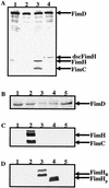
FimH has sufficient information for FimD binding. (A) Western blot developed with anti-FimCH and anti-FimD antiserum of outer membrane preparations after nickel chromatography from the strains C600/pETS7/pMMB66 (FimDHis) (lane 1), C600/pETS7/pMMB-FimH (FimDHis + FimH) (lane 2), C600/pETS7/pETS1007 (FimDHis + FimC + FimH) (lane 3), and C600/pETS7/pMMB-dscFimH (FimDHis + dscFimH) (lane 4). (B to D) Coomassie blue-stained SDS-polyacrylamide gel (B), Western blot developed with anti-FimCH antiserum (C), and Western blot developed with anti-HA antiserum (D) of outer membrane preparations after nickel chromatography from the strains C600/pCD/pBad18-Kn (FimC + FimDHis) (lanes 1), C600/pCD/pBad-FimH (FimC + FimDHis + FimH) (lanes 2), C600/pCD/p601 (FimC + FimDHis + FimHR) (lanes 3), C600/pCD/p421 (FimC + FimDHis + FimHP) (lanes 4), and C600/pCD/pMC10 (FimC + FimDHis + CsgE-HA) (lanes 5).
FIG. 3.
FIG. 3.
dscFimG is not incorporated into a growing pilus. (A) Coomassie blue-stained gel of FimCHis-FimG-FimH complexes incubated at 95°C (lane 1) or 25°C (lane 2), purified dscFimG (lane 3), or dscFimG-FimH complexes incubated at 95°C (lane 4) or 25°C (lane 5). (B to D) Coomassie blue-stained gel (B), Western blot developed with anti-FimCH antiserum (C), or Western blot developed with anti-FimG antiserum (D) of outer membrane preparations after nickel chromatography from the strains C600/pCD-Kn/pMB7/pTrc99A (FimC + FimDHis + FimH) (lanes 1), C600/CD-Kn/pMB7/pTrc-FimG (FimC + FimDHis + FimH + FimG) (lanes 2), and C600/pCD-Kn/pMB7/pTrc-dscFimG (FimC + FimDHis + FimH + dscFimG) (lanes 3). The ability of the strains to hemagglutinate (HA) guinea pig red blood cells is indicated at the bottom of panel D (+, HA; −, no HA).
FIG. 3.
FIG. 3.
dscFimG is not incorporated into a growing pilus. (A) Coomassie blue-stained gel of FimCHis-FimG-FimH complexes incubated at 95°C (lane 1) or 25°C (lane 2), purified dscFimG (lane 3), or dscFimG-FimH complexes incubated at 95°C (lane 4) or 25°C (lane 5). (B to D) Coomassie blue-stained gel (B), Western blot developed with anti-FimCH antiserum (C), or Western blot developed with anti-FimG antiserum (D) of outer membrane preparations after nickel chromatography from the strains C600/pCD-Kn/pMB7/pTrc99A (FimC + FimDHis + FimH) (lanes 1), C600/CD-Kn/pMB7/pTrc-FimG (FimC + FimDHis + FimH + FimG) (lanes 2), and C600/pCD-Kn/pMB7/pTrc-dscFimG (FimC + FimDHis + FimH + dscFimG) (lanes 3). The ability of the strains to hemagglutinate (HA) guinea pig red blood cells is indicated at the bottom of panel D (+, HA; −, no HA).
FIG. 4.
FIG. 4.
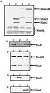
FimGH is stable in the periplasm and is targeted for pilus assembly. (A) Western blot of periplasms prepared from strains C600/pMMB-FimH (FimH) (lane 1), C600/pHJ9205/pMMB-FimH (FimC + FimH) (lane 2), C600/pMMB-FimGH (FimGH) (lane 3), and C600/pHJ9205/pMMB-FimGH (FimC + FimGH) (lane 4). The Western blot was developed with anti-FimCH antiserum. (B to E) Coomassie blue-stained SDS-polyacrylamide gel (B), Western blot developed with anti-FimCH antiserum (C), Western blot developed with anti-FimG antiserum (D), or Western blot developed with anti-FimF antiserum (E) of outer membrane preparations after nickel chromatography from strains C600/pCD-Kn/pMB10/pTrc99A (FimC + FimDHis + FimGH) (lanes 1), C600/pCD-Kn/pMB10/pTrc-FimG (FimC + FimDHis + FimGH + FimG) (lanes 2), and C600/pCD-Kn/pMB10/pTrc-FimF (FimC + FimDHis + FimGH + FimF) (lanes 3).
FIG. 5.
FIG. 5.
Model of type 1 pilus assembly at the usher. The FimC-FimH complex binds to the FimD usher, inducing a conformation change in FimD (42), presumably into an assembly-competent state. We hypothesize that the FimH receptor binding domain (HR) is inserted into the FimD pore, allowing the FimH pilin domain (HP) to be exposed on the periplasmic side of FimD to remain available for donor strand exchange with FimC-FimG complexes. dscFimG was not incorporated into the growing pilus, suggesting that the chaperone is required for appropriate targeting and assembly of subunits into the pilus. The black line on FimC represents the FimC G1β strand completing the Ig fold of either FimG or FimH. The yellow line of dscFimG represents the N-terminal extension of FimF completing the Ig fold of FimG.

Similar articles

Cited by

References

    1. Abraham, S. N., J. D. Goguen, D. Sun, P. Klemm, and E. H. Beachey. 1987. Identification of two ancillary subunits of Escherichia coli type 1 fimbriae by using antibodies against synthetic oligopeptides of fim gene products. J. Bacteriol. 169:5530-5536. - PMC - PubMed
    1. Amann, E., B. Ochs, and K. J. Abel. 1988. Tightly regulated tac promoter vectors useful for the expression of unfused and fused proteins in Escherichia coli. Gene 69:301-315. - PubMed
    1. Barnhart, M. M., J. S. Pinkner, G. E. Soto, F. G. Sauer, S. Langermann, G. Waksman, C. Frieden, and S. J. Hultgren. 2000. PapD-like chaperones provide the missing information for folding of pilin proteins. Proc. Natl. Acad. Sci. USA 97:7709-7714. - PMC - PubMed
    1. Batchelor, M., S. Prasannan, S. Daniell, S. Reece, I. Connerton, G. Bloomberg, G. Dougan, G. Frankel, and S. Matthews. 2000. Structural basis for recognition of the translocated intimin receptor (Tir) by intimin from enteropathogenic Escherichia coli. EMBO J. 19:2452-2464. - PMC - PubMed
    1. Bock, K., M. E. Breimer, A. Brignole, G. C. Hansson, K.-A. Karlsson, G. Larson, H. Leffler, B. E. Samuelsson, N. Strömberg, C. Svanborg-Edén, and J. Thurin. 1985. Specificity of binding of a strain of uropathogenic Escherichia coli to Galα(1-4)Gal-containing glycosphingolipids. J. Biol. Chem. 260:8545-8551. - PubMed

Publication types

MeSH terms