Comparative Study
. 2003 May;185(9):2848-55.
doi: 10.1128/JB.185.9.2848-2855.2003.

Substrate specificity of the nonribosomal peptide synthetase PvdD from Pseudomonas aeruginosa

Affiliations
Comparative Study

Substrate specificity of the nonribosomal peptide synthetase PvdD from Pseudomonas aeruginosa

David F Ackerley et al. J Bacteriol. 2003 May.

Abstract

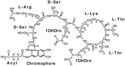
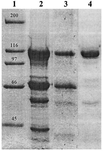
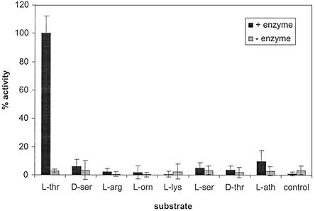
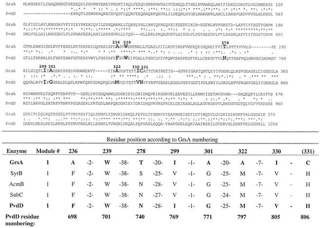
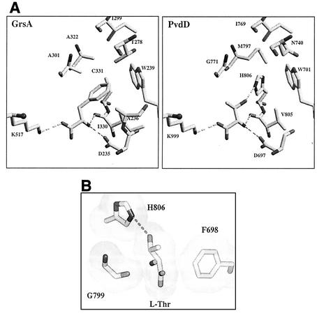
Pseudomonas aeruginosa PAO1 secretes a siderophore, pyoverdine(PAO), which contains a short peptide attached to a dihydroxyquinoline moiety. Synthesis of this peptide is thought to be catalyzed by nonribosomal peptide synthetases, one of which is encoded by the pvdD gene. The first module of pvdD was overexpressed in Escherichia coli, and the protein product was purified. L-Threonine, one of the amino acid residues in pyoverdine(PAO), was an effective substrate for the recombinant protein in ATP-PP(i) exchange assays, showing that PvdD has peptide synthetase activity. Other amino acids, including D-threonine, L-serine, and L-allo-threonine, were not effective substrates, indicating that PvdD has a high degree of substrate specificity. A three-dimensional modeling approach enabled us to identify amino acids that are likely to be critical in determining the substrate specificity of PvdD and to explore the likely basis of the high substrate selectivity. The approach described here may be useful for analysis of other peptide synthetases.


Figures

FIG. 1.
FIG. 1.
Structure of pyoverdinePAO produced by P. aeruginosa PAO1 (1). The positions of amino acid residues, the dihydroxyquinoline chromophore, and an acyl group are shown. The acyl group can be a succinyl, α-ketoglutaryl, or succinamide residue. fOHOrn, l-N5-formyl-N5-hydroxyornithine.
FIG. 2.
FIG. 2.
Affinity purification of His-PvdD. E. coli BL21(pPROEX::mod1) was grown at 18°C in LB containing betaine and sorbitol and was induced to express His-PvdD as described in the text. The cells were sonicated, and His-PvdD was purified by using a nickel affinity column. Protein preparations obtained following a single passage (lane 2), following two passages (lane 3), and following an ATP incubation step (lane 4) were analyzed by sodium dodecyl sulfate-polyacrylamide gel electrophoresis. Lane 1 contained Bio-Rad broad-range standard markers.
FIG. 3.
FIG. 3.
ATP-PPi exchange activity of His-PvdD. ATP-PPi exchange activity was measured with different amino acids. The highest exchange activity observed was defined as 100%. The error bars indicate one standard deviation. L-thr, l-threonine; D-ser, d-serine; L-arg, l-arginine; L-orn, l-ornithine; L-lys, l-lysine; L-ser, l-serine; D-thr, d-threonine; L-ath, l-allo-threonine.
FIG. 4.
FIG. 4.
Identification of substrate-determining residues of PvdD. The sequence of the A domain of the first module of PvdD was aligned with that of GrsA. An asterisk indicates identical residues, a colon indicates residues with a high level of chemical similarity, and a period indicates residues with a lower level of chemical similarity. The eight residues of PvdD that are predicted to determine the amino acid substrate specificity (the coding residues [5, 38]) are indicated by boldface type in the alignment. Residue 331 (GrsA numbering) was considered to be a coding residue by Stachelhaus et al. (38) but not by Challis et al. (5). The predicted coding residues of three other l-threonine-activating A domains are from SyrB, a syringomycin synthetase from Pseudomonas syringae (15); AcmB, an actinomycin synthetase from Streptomyces chrysomallus (34); and SnbC, a pristinamycin synthetase from Streptomyces pristinaespiralis (42). Threonine activation was demonstrated by ATP-PPi exchange for SnbC and SyrB. For AcmB, assignment of threonine as the substrate was inferred from the linear order of the modules in the chromosome according to the nearly invariant colinearity rule (reviewed in reference 24).
FIG. 5.
FIG. 5.
Model explaining the substrate specificity of PvdD. (A) Phenylalanine-binding site of GrsA (8) and predicted threonine-binding site of PvdD. Carbon and sulfur atoms are represented by light grey bars, and oxygen and nitrogen are represented by dark grey bars. The view is from the same angle in both cases, and the substrate amino acids are in the middle of each frame. All 10 residues that line the amino acid substrate-binding pocket are shown in the GrsA structure, but residue F698 has been removed from the foreground of the PvdD structure for clarity. The cosubstrate AMP molecule is predicted to occupy much of the left side of each frame. Three hydrogen bonds that are predicted to stabilize the substrate amino and carboxyl groups in the active sites are indicated by dashed lines. A substrate-determining hydrogen bond is also predicted to be formed between the threonine side chain hydroxyl and a nitrogen atom in the side chain of H806 of PvdD. (B) View rotated 90o with respect to the view shown in panel A. The van der Waals surfaces of l-threonine, G799, and the side chains of F698 and H806 of PvdD are shown. The substrate l-threonine is predicted to be sandwiched between F698 and the main-chain carbonyl of G799. Modeling of insertion of other isomers of threonine (d-threonine and l-allo-threonine) into the predicted PvdD substrate-binding pocket showed that while these isomers could form hydrogen bonds with D697, V805, and K999 (as shown for l-threonine in panel A), they would be unable to form a hydrogen bond with H806 without being unacceptably close to F698 or G799 (data not shown).

References

    1. Abdallah, M. A. 1991. Pyoverdines and pseudobactins, p. 139-153. In G. Winkelmann (ed.), CRC handbook of microbial iron chelates. CRC Press, Boca Raton, Fla.
    1. Barth, S., M. Huhn, B. Matthey, A. Klimka, E. A. Galinski, and A. Engert. 2000. Compatible-solute-supported periplasmic expression of functional recombinant proteins under stress conditions. Appl. Environ. Microbiol. 66:1572-1579. - PMC - PubMed
    1. Blackwell, J. R., and R. Horgan. 1991. A novel strategy for production of a highly expressed recombinant protein in an active form. FEBS Lett. 295:10-12. - PubMed
    1. Buchberger, A., and B. Bukau. 1997. Escherichia coli DnaK, p. 22-24. In M. J. Gething (ed.), Guidebook to molecular chaperones and protein-folding catalysts. Sambrook and Tooze, Oxford University Press, Oxford, United Kingdom.
    1. Challis, G. L., J. Ravel, and C. A. Townsend. 2000. Predictive, structure-based model of amino acid recognition by nonribosomal peptide synthetase adenylation domains. Chem. Biol. 7:211-224. - PubMed

Publication types

MeSH terms

LinkOut - more resources