. 2003 May;185(9):2910-9.
doi: 10.1128/JB.185.9.2910-2919.2003.

The YSIRK-G/S motif of staphylococcal protein A and its role in efficiency of signal peptide processing

Affiliations

The YSIRK-G/S motif of staphylococcal protein A and its role in efficiency of signal peptide processing

Taeok Bae et al. J Bacteriol. 2003 May.

Abstract

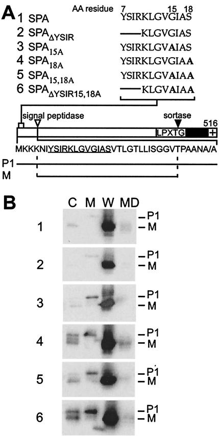
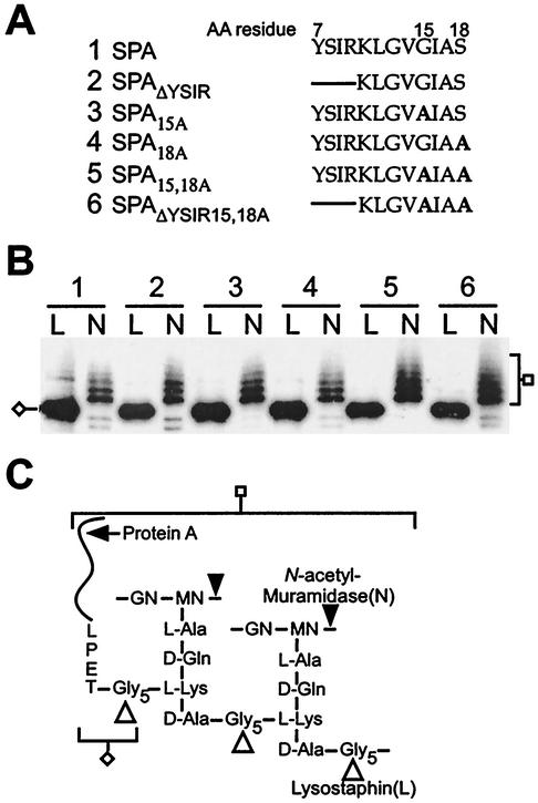
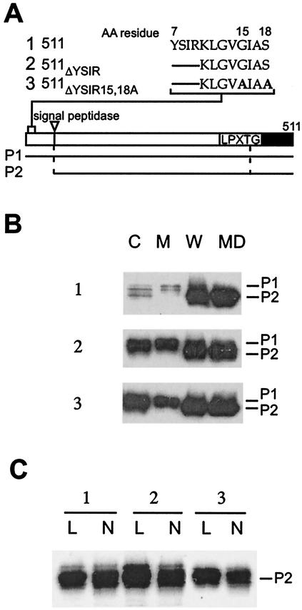
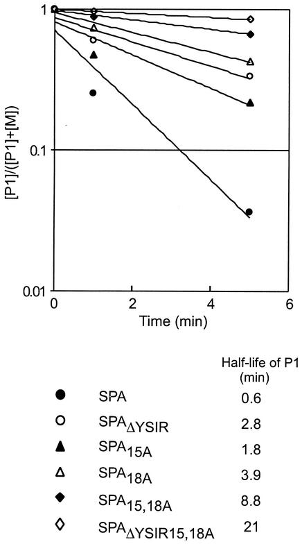
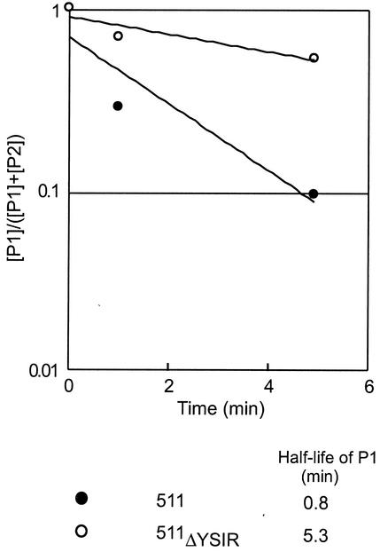
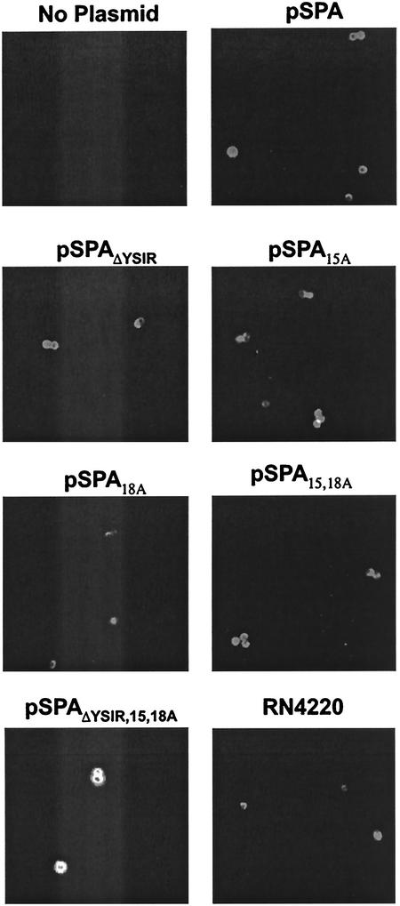
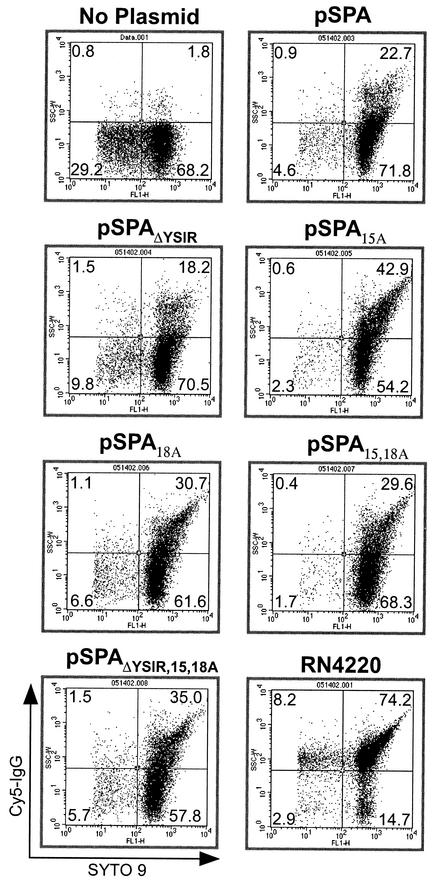
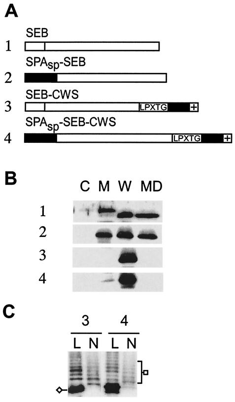
Many surface proteins of pathogenic gram-positive bacteria are linked to the cell wall envelope by a mechanism requiring a C-terminal sorting signal with an LPXTG motif. Surface proteins of Streptococcus pneumoniae harbor another motif, YSIRK-G/S, which is positioned within signal peptides. The signal peptides of some, but not all, of the 20 surface proteins of Staphylococcus aureus carry a YSIRK-G/S motif, whereas those of surface proteins of Listeria monocytogenes and Bacillus anthracis do not. To determine whether the YSIRK-G/S motif is required for the secretion or cell wall anchoring of surface proteins, we analyzed variants of staphylococcal protein A, an immunoglobulin binding protein with an LPXTG sorting signal. Deletion of the YSIR sequence or replacement of G or S significantly reduced the rate of signal peptide processing of protein A precursors. In contrast, cell wall anchoring or the functional display of protein A was not affected. The fusion of cell wall sorting signals to reporter proteins bearing N-terminal signal peptides with or without the YSIRK-G/S motif resulted in hybrid proteins that were anchored in a manner similar to that of wild-type protein A. The requirement of the YSIRK-G/S motif for efficient secretion implies the existence of a specialized mode of substrate recognition by the secretion pathway of gram-positive cocci. It seems, however, that this mechanism is not essential for surface protein anchoring to the cell wall envelope.


Figures

FIG. 1.
FIG. 1.
Cellular locations of protein A mutants. (A) The diagram displays wild-type protein A (SPA) with the N-terminal signal peptide, the LPXTG motif, the C-terminal hydrophobic domain (black bar), and the charged tail (+). The amino acid sequence of the signal peptide is shown below the diagram, in which the YSIRK-G/S sequence is underlined. The mutations in the YSIRK-G/S motif are shown above the diagram. The lines in the sequences indicate a deletion of amino acids (AA), while boldfaced A's indicate alanine substitutions. The precursor (P1) and mature species of protein A (M) are indicated below the diagram. (B) Cell fractionation of S. aureus OS2 expressing protein A mutants. Bacteria were grown to mid-log phase, and the cultures were fractionated into medium (MD), cell wall (W), membrane (M), and cytoplasmic (C) compartments. TCA-precipitated samples were subjected to SDS-10% PAGE, and protein A was detected by immunoblotting with monoclonal antibody.
FIG. 2.
FIG. 2.
Anchoring of protein A mutants to the cell wall. (A) YSIRK-G/S motif and mutant sequences of protein A and its variants. The lines in the sequences indicate a deletion of amino acids (AA), while boldfaced A's represent alanine substitutions. (B) S. aureus OS2 cells expressing protein A mutants were treated with either lysostaphin (L) or N-acetylmuramidase (N). The digested cell wall components were subjected to SDS-10% PAGE, and protein A mutants were detected by immunoblotting with monoclonal antibody. Cleavage products of specific enzymes are indicated. (C) Structure of the cell wall of S. aureus and cleavage sites for lysostaphin and N-acetylmuramidase. Protein A anchored to the peptidoglycan is drawn as a curved line, and the LPET sequence is tethered to the pentaglycine cell wall crossbridge (cleaved and anchored LPXTG motif).
FIG. 3.
FIG. 3.
YSIRK-G/S motif mutants of C-terminally truncated protein A. (A) Diagram of the truncated protein A and sequences of YSIRK-G/S motif mutants. The diagram shows the cleavage site for signal peptidase as well as the LPXTG motif and the C-terminal hydrophobic domain (black bar). The P1 and P2 precursors (cleavage products of signal peptidase) are shown below the diagram. AA, amino acid. (B) S. aureus OS2 cells expressing protein A variants were grown to mid-log phase, and cultures were fractionated into medium (MD), cell wall (W), membrane (M), and cytoplasmic (C) compartments. The samples were subjected to SDS-10% PAGE, and protein A was detected by immunoblotting with monoclonal antibody. The migration of P1 and P2 precursors is indicated. (C) S. aureus OS2 cells expressing protein A mutants were treated with either lysostaphin (L) or N-acetylmuramidase (N). The digested cell wall components were subjected to SDS-10% PAGE, and protein A was detected by immunoblotting with monoclonal antibody.
FIG. 4.
FIG. 4.
Precursor processing of YSIRK-G/S mutants of protein A. S. aureus cultures were pulse-labeled with [35S]methionine, and labeling was quenched with nonradioactive methionine. Aliquots of the culture were precipitated with TCA to stop protein processing at 0, 1, or 5 min after the conclusion of the pulse. Staphylococci were digested with lysostaphin, and proteins were again precipitated with TCA. Proteins were solubilized in hot SDS, immunoprecipitated with polyclonal antibody, and subjected to SDS-10% PAGE and PhosphorImager analysis. The logarithmic ratio of the concentration of the P1 precursor ([P1]) to the sum of the P1 precursor and mature protein A ([P1] + [M]) was calculated and plotted against time. The half-life of each P1 species was calculated by using regression curves.
FIG. 5.
FIG. 5.
Precursor processing of a YSIRK-G/S mutant of C-terminally truncated protein A. S. aureus was pulse-labeled with [35S]methionine, and the labeling was quenched with nonradioactive methionine. Aliquots of the culture were precipitated with TCA to stop protein processing 0, 1, or 5 min after the pulse. Staphylococci were digested with lysostaphin, and proteins were again precipitated with TCA. Proteins were solubilized in hot SDS, immunoprecipitated with polyclonal antibody, and subjected to SDS-10% PAGE and PhosphorImager analysis. Because C-terminally truncated protein A is not subject to sortase-mediated cleavage and anchoring at the LPXTG motif, signal peptide processing generates only the P2 precursor. The logarithmic ratio of the concentration of the P1 precursor ([P1]) to the sum of the P1 precursor and the P2 precursor of protein A ([P1] + [P2]) was calculated and plotted against time. The half-life of each P1 species was calculated by using regression curves.
FIG. 6.
FIG. 6.
Surface display of protein A mutants as examined by immunofluorescence microscopy. Cells were grown to mid-log phase, and surface-displayed protein A was stained with Cy5-conjugated goat IgG. Samples were viewed by fluorescence microscopy, and images were captured with a charge-coupled-device camera. The S. aureus RN4220 strain (spa+) was used as a positive control. Note that all plasmids are transformed into strain OS2 (spa::ermC), which does not express protein A.
FIG. 7.
FIG. 7.
Measurements of the surface display of protein A in staphylococcal populations by FACS analysis. Staphylococci were grown to mid-log phase, stained with Cy5-conjugated goat IgG and SYTO 9, and analyzed with a FACS. The S. aureus RN4220 strain (spa+) was used as positive control, while the OS2 strain (spa::ermC) (no plasmid) was used as a negative control. Note that all plasmids are transformed into strain OS2 (spa::ermC), which does not express protein A.
FIG. 8.
FIG. 8.
Cell wall sorting of enterotoxin B fusions. (A) The diagram displays SEB and SPASP-SEB fusion proteins. The signal peptide of SEB does not harbor the YSIRK-G/S motif and is represented by a white block. SPASP does encompass the YSIRK-G/S motif and is shown as a gray block. The LPXTG motif, the C-terminal hydrophobic domain (black bar), and the charged tail (+) of SPA are indicated in the drawing. (B) Cellular locations of the SPA-SEB fusion proteins. Cells were grown to mid-log phase and fractionated into medium (MD), cell wall (W), membrane (M), and cytoplasm (C) compartments. The samples were subjected to SDS-12% PAGE and analyzed by immunoblotting with polyclonal anti-SEB antibody. (C) Cells at mid-log phase were treated with either lysostaphin (L) or N-acetylmuramidase (N) in the presence of 0.5 M sucrose. The digested cell wall components were collected, subjected to SDS-12% PAGE, and then analyzed by immunoblotting with anti-SEB antibody.

References

    1. Bassford, P. J., Jr., T. J. Silhavy, and J. Beckwith. 1979. Use of gene fusion to study secretion of maltose-binding protein into Escherichia coli periplasm. J. Bacteriol. 139:19-31. - PMC - PubMed
    1. Beck, K., L.-F. Wu, J. Brunner, and M. Müller. 2000. Discrimination of SRP- and SecA/SecB-dependent substrates involves selective recognition of nascent chains by SRP and trigger factor. EMBO J. 19:134-143. - PMC - PubMed
    1. Benson, S. A., M. N. Hall, and T. J. Silhavy. 1985. Genetic analysis of protein export in Escherichia coli K12. Annu. Rev. Biochem. 54:101-134. - PubMed
    1. Bernstein, H. D., M. A. Poritz, K. Strub, P. J. Hoben, S. Brenner, and P. Walter. 1989. Model for signal sequence recognition from amino-acid sequence of 54K subunit of signal recognition particle. Nature 340:482-486. - PubMed
    1. Blobel, G. 1980. Intracellular protein topogenesis. Proc. Natl. Acad. Sci. USA 77:1496-1500. - PMC - PubMed

Publication types

MeSH terms

LinkOut - more resources