. 2003 Apr 29;100(9):5296-301.
doi: 10.1073/pnas.0831002100. Epub 2003 Apr 16.

A 1-Mb resolution radiation hybrid map of the canine genome

Affiliations

A 1-Mb resolution radiation hybrid map of the canine genome

Richard Guyon et al. Proc Natl Acad Sci U S A. .

Abstract

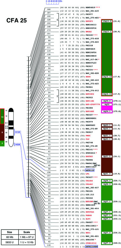
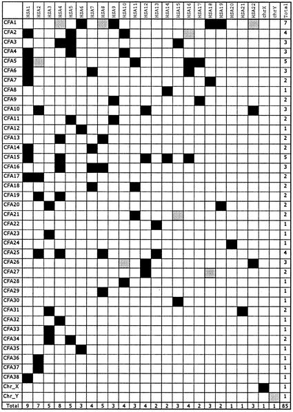
The purebred dog population consists of >300 partially inbred genetic isolates or breeds. Restriction of gene flow between breeds, together with strong selection for traits, has led to the establishment of a unique resource for dissecting the genetic basis of simple and complex mammalian traits. Toward this end, we present a comprehensive radiation hybrid map of the canine genome composed of 3,270 markers including 1,596 microsatellite-based markers, 900 cloned gene sequences and ESTs, 668 canine-specific bacterial artificial chromosome (BAC) ends, and 106 sequence-tagged sites. The map was constructed by using the RHDF5000-2 whole-genome radiation hybrid panel and computed by using MULTIMAP and TSP/CONCORDE. The 3,270 markers map to 3,021 unique positions and define an average intermarker distance corresponding to 1 Mb. We also define a minimal screening set of 325 highly informative well spaced markers, to be used in the initiation of genome-wide scans. The well defined synteny between the dog and human genomes, established in part as a function of this work by the identification of 85 conserved fragments, will allow follow-up of initial findings of linkage by selection of candidate genes from the human genome sequence. This work continues to define the canine system as the method of choice in the pursuit of the genes causing mammalian variation and disease.


Figures

Figure 1
Figure 1
RH map of CFA25. The position of each marker is reported along the RH map, symbolized by a vertical bar. The RH map shows the five maps generated by TSP/CONCORDE. Maps are highlighted by horizontal bars of variable lengths. When a marker is present on all five maps at the same position, the horizontal bar has a maximum length indicating high confidence; shorter bars reflect a lower confidence level. The scale of 0–100% reflects the relative confidence level. Marker number, as it appears in the consensus map, is indicated in parentheses. In scrambled regions, markers occupying several positions are bracketed to narrow the problematic region into smaller intervals. Marker names indicated in red correspond to gene-based markers (type I); other markers are black (see Table 1 for nomenclature). MSS-2 markers have three asterisks; polymorphic STS, genes, or BAC ends have one. Colored boxes to the right of the markers display human segments with the chromosomal band position. The corresponding position in nucleotide (Mb) of human putative orthologs is indicated between brackets. Data are based on NCBI Build 31. At the left of the RH map, a 4′,6-diamidino-2-phenylindole-banded ideogram is drawn. Markers assigned to chromosomes by FISH are linked to their RH map positions by colored lines (11). Colored bars correspond to the human evolutionary CS. Numbers indicate HSA origin as determined by reciprocal chromosome painting (30, 31). Distances between RH markers are reported in TSP units between horizontal bars. The physical size of each chromosome (in Mb), as determined by flow sorting (25), and the RH group total size (in TSP units) are reported in the frame. The correspondences between TSP unit and kb are also reported in the frame. Figures for all chromosomes are available at www-recomgen.univ-rennes1.fr/doggy.html and www.fhcrc.org/science/dog_genome/dog.html.
Figure 2
Figure 2
Schematic view of RH conserved segments and singletons between dog and human. CS between both species are illustrated by black squares; singletons are illustrated by gray squares. For each CFA and HSA, the total number of CS is reported in the last column and the last line, respectively.

References

    1. Ostrander E A, Giniger E. Am J Hum Genet. 1997;61:475–480. - PMC - PubMed
    1. Ostrander E A, Galibert F, Patterson D F. Trends Genet. 2000;16:117–123. - PubMed
    1. Patterson D. J Vet Intern Med. 2000;14:1–9. - PubMed
    1. Wilcox B, Walkowicz C. Atlas of Dog Breeds of the World. Neptune City, NJ: T.H.F.; 1995.
    1. Ostrander E A, Kruglyak L. Genome Res. 2000;10:1271–1274. - PubMed

Publication types

LinkOut - more resources