

Hypoxia inhibits contraction but not calcium channel currents or changes in intracellular calcium in arteriolar muscle cells
- PMID: 12700582
- PMCID: PMC1382023
- DOI: 10.1038/sj.mn.7800178
Hypoxia inhibits contraction but not calcium channel currents or changes in intracellular calcium in arteriolar muscle cells
Abstract
Objective: We tested the hypothesis that hypoxia inhibits currents through L-type Ca(2+) channels and inhibits norepinephrine-induced rises in intracellular Ca(2+) in cremasteric arteriolar muscle cells, thus accounting for the inhibitory effect of hypoxia on norepinephrine-induced contraction of these cells.
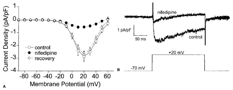
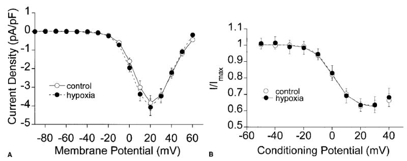
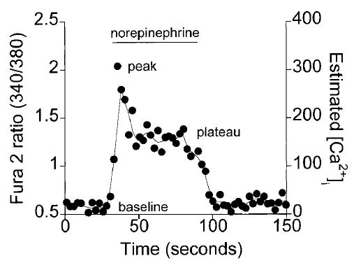
Methods: Single smooth muscle cells were enzymatically isolated from second-order and third-order arterioles from hamster cremaster muscles. The effects of hypoxia (partial pressure of oxygen: 10-15 mm Hg) were examined on Ba(2+) (10 mM) currents through L-type Ca(2+) channels by use of the perforated patch clamp technique. Also, the effect of hypoxia on norepinephrine-induced calcium changes was studied using Fura 2 microfluorimetry.
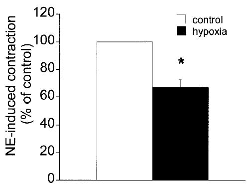
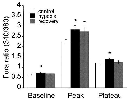
Results: Hypoxia inhibited the norepinephrine-induced (10 microM) contraction of single arteriolar muscle cells by 32.9 +/- 5.6% (mean +/- SE, n = 4). However, hypoxia had no significant effect on whole-cell currents through L-type Ca(2+) channels: the peak current densities measured at +20 mV were -3.83 +/- 0.40 pA/pF before hypoxia and -3.97 +/- 0.36 pA/pF during hypoxia (n = 15; p > 0.05). In addition, hypoxia did not inhibit Ca(2+) transients in arteriolar muscle cells elicited by 10 microM norepinephrine. Instead, hypoxia increased basal Ca(2+) (13.8 +/- 3.2%) and augmented peak Ca(2+) levels (29.4 +/- 7.3%) and steady-state Ca(2+) levels (15.2 +/- 5.4%) elicited by 10 microM norepinephrine (n = 21; p < 0.05).
Conclusions: These data indicate that hypoxia inhibits norepinephrine-induced contraction of single cremasteric arteriolar muscle cells by a mechanism that involves neither L-type Ca(2+) channels nor norepinephrine-induced Ca(2+) mobilization. Instead, our findings suggest that hypoxia must inhibit norepinephrine-induced contraction by affecting a component of the signaling pathway that lies downstream from the increases in Ca(2+) produced by this neurotransmitter.
Figures
References
-
- Bongard O, Bounameaux H, Fagrell B. Effects of oxygen inhalation on skin microcirculation in patients with peripheral arterial occlusive disease. Circulation. 1992;86:878–886. - PubMed
-
- Bredle DL, Bradley WE, Chapler CK, Cain SM. Muscle perfusion and oxygenation during local hyperoxia. J Appl Physiol. 1988;65:2057–2062. - PubMed
-
- Franco-Obregon A, Lopez-Barneo J. Low PO2 inhibits calcium channel activity in arterial smooth muscle cells. Am J Physiol. 1996;271:H2290–H2299. - PubMed
Publication types
MeSH terms
Substances
Grants and funding
LinkOut - more resources
Full Text Sources
Miscellaneous
