Review
. 2003;4(4):212.
doi: 10.1186/gb-2003-4-4-212. Epub 2003 Apr 1.

Protein prenyltransferases

Affiliations
Review

Protein prenyltransferases

Sebastian Maurer-Stroh et al. Genome Biol. 2003.

Abstract

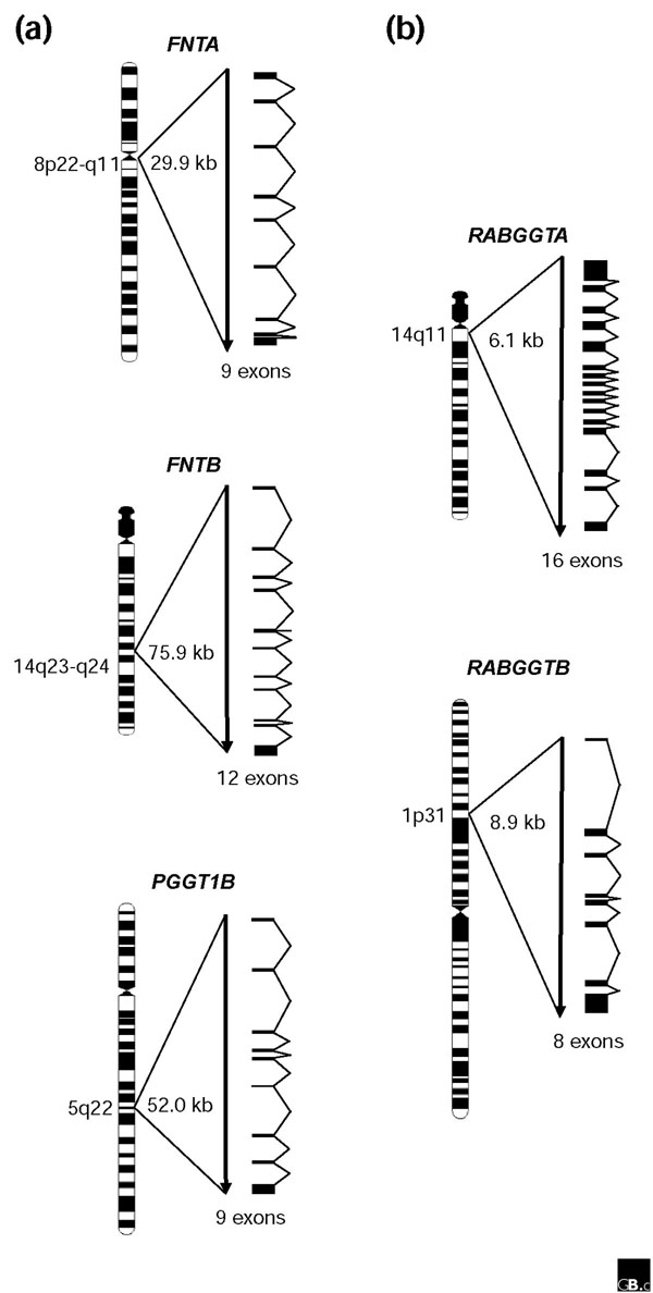
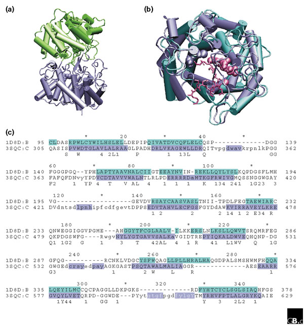
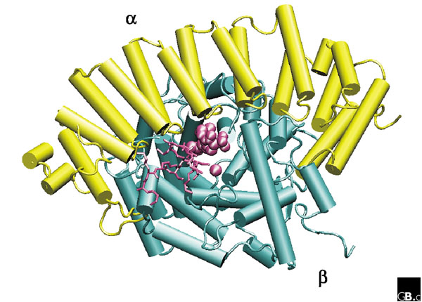
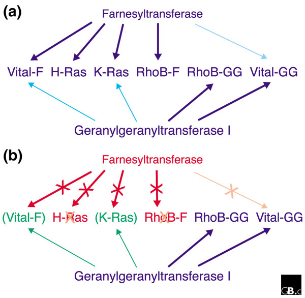
Three different protein prenyltransferases (farnesyltransferase and geranylgeranyltransferases I and II) catalyze the attachment of prenyl lipid anchors 15 or 20 carbons long to the carboxyl termini of a variety of eukaryotic proteins. Farnesyltransferase and geranylgeranyltransferase I both recognize a 'Ca1a2X' motif on their protein substrates; geranylgeranyltransferase II recognizes a different, non-CaaX motif. Each enzyme has two subunits. The genes encoding CaaX protein prenyltransferases are considerably longer than those encoding non-CaaX subunits, as a result of longer introns. Alternative splice forms are predicted to occur, but the extent to which each splice form is translated and the functions of the different resulting isoforms remain to be established. Farnesyltransferase-inhibitor drugs have been developed as anti-cancer agents and may also be able to treat several other diseases. The effects of these inhibitors are complicated, however, by the overlapping substrate specificities of geranylgeranyltransferase I and farnesyltransferase.


Figures

Figure 1
Figure 1
Gene structures and chromosomal locations of human protein prenyltransferase subunit genes. The chromosome diagrams and the locations of the genes on the cytogenetic map are according to the NCBI MapViewer [67]. The sizes of the genes are indicated but are not drawn to scale. (a) Genes encoding CaaX protein prenyltransferases are relatively long; (b) genes encoding non-CaaX protein prenyltransferases are much shorter.
Figure 2
Figure 2
The structural relationship between protein prenyltransferase β subunits and squalene-hopene cyclases. (a) The complete structure of squalene-hopene cyclase from the bacterium Alicyclobacillus acidocaldarius (Protein Data Bank (PDB) identifier 3SQC [68]). (b) Structural superposition of the conserved (α-α)6 barrels of squalene-hopene cyclase (blue) and the rat FT β subunit (cyan; see Figure 3 for the full structure of the FT). Blue spheres, functionally important residues in the half of the squalene-hopene cyclase that is shown (the sphere indicates the van der Waals radius); pink spheres, farnesyl-pyrophosphate and Zn2+ bound to the β subunit of FT; pink residues in stick representation, the Ca1a2X peptide of the substrate protein. (c) Alignment of the two proteins on the basis of the structural superposition. Secondary structural elements are colored as in (b); black residues with colored background represent α helix and white residues with colored background represent β sheet. Conserved residues and similar residues are shown below the alignment; 1, polar; 2, small; 3, aromatic; 4, hydrophobic. The lower-case letters represent residues within 3SQC that were not easily superimposed on the 1D8D structure (that is, gaps in 1D8D). The molecular representations were created using VMD [69].
Figure 3
Figure 3
The complete structure of rat FT (PDB identifier 1D8D [70]). The spheres in the center represent the farnesyl-pyrophosphate and Zn2+, and the amino acids in stick representation are the CaaX motif of the protein substrate. The molecular representation was created using VMD [69].
Figure 4
Figure 4
Differential prenylation of substrates of CaaX protein prenyltransferases (a) under normal conditions and (b) in the presence of a FT inhibitor. Substrates are shown in the middle of each panel, with the two enzymes above and below. 'Vital-F' and 'Vital-GG' represent all the proteins for which farnesylation (Vital-F) or geranylgeranylation (Vital-GG) are essential; RhoB-GG represents geranylgeranylated RhoB and RhoB-F represents farnesylated RhoB. Thick arrow, normal prenylation activity; thin arrow, reduced prenylation activity through cross-specificity of the non-preferred enzyme; arrows with crosses, blocked prenylation activity during inhibition of FT; brackets, substituting prenylation activity by GGT1 during inhibition of FT.

References

    1. Casey PJ, Seabra MC. Protein prenyltransferases. J Biol Chem. 1996;271:5289–5292. A short minireview describing protein prenyltransferases, written before the structure was known. - PubMed
    1. AceView http://www.humangenes.org Alignment of expressed sequence tags and mRNAs to the human genome, showing alternative splice forms.
    1. Andres DA, Milatovich A, Ozcelik T, Wenzlau JM, Brown MS, Goldstein JL, Francke U. cDNA cloning of the two subunits of human CAAX farnesyltransferase and chromosomal mapping of FNTA and FNTB loci and related sequences. Genomics. 1993;18:105–112. Cloning of human FNTA and FNTB; 'related sequences' refers to processed pseudogenes. - PubMed
    1. Dhawan P, Yang E, Kumar A, Mehta KD. Genetic complexity of the human geranylgeranyltransferase I beta-subunit gene: a multigene family of pseudogenes derived from mis-spliced transcripts. Gene. 1998;210:9–15. The authors suggest that there are 13 GGT1B pseudogenes, but these seem to correspond to only two in the human genome. - PubMed
    1. Blatch GL, Lassle M. The tetratricopeptide repeat: a structural motif mediating protein-protein interactions. BioEssays. 1999;21:932–939. Review describing the family of TPR-containing proteins. - PubMed

Publication types

Substances

LinkOut - more resources