Comparative Study
. 2003 Apr 17:3:2.
doi: 10.1186/1472-6793-3-2. Epub 2003 Apr 17.

Is there evidence of fetal-maternal heart rate synchronization?

Affiliations
Comparative Study

Is there evidence of fetal-maternal heart rate synchronization?

Peter Van Leeuwen et al. BMC Physiol. .

Abstract

Background: The prenatal condition offers a unique possibility of examining physiological interaction between individuals. Goal of this work was to look for evidence of coordination between fetal and maternal cardiac systems.

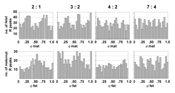
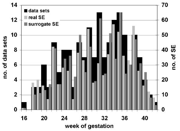
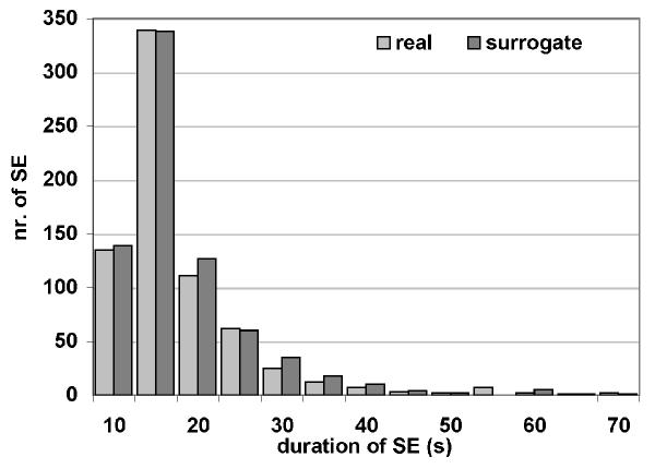
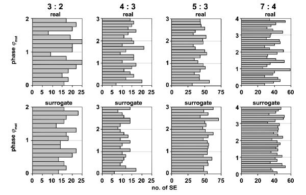
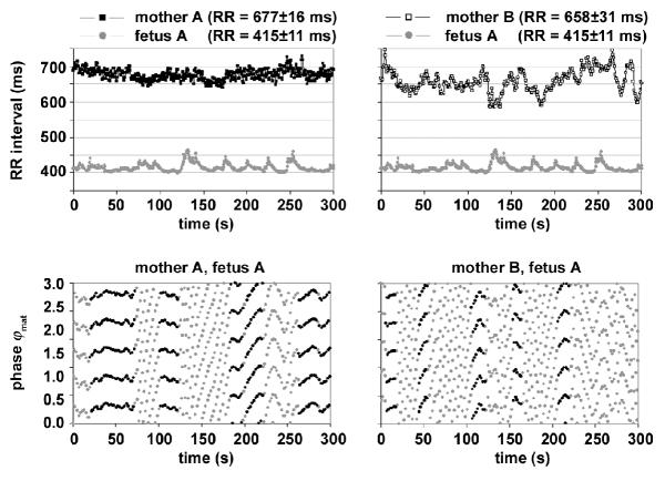
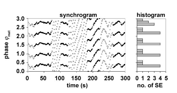
Methods: 177 magnetocardiograms were recorded in 62 pregnancies (16th-42nd week of gestation). Fetal and maternal RR interval time series were constructed and the phases, i.e. the timing of the R peaks of one time series in relation to each RR interval of the other were determined. The distributions of these phases were examined and synchrograms were constructed for real and surrogate pairs of fetal and maternal data sets. Synchronization epochs were determined for defined n:m coupling ratios.

Results: Differences between real and surrogate data could not be found with respect to number of synchronization epochs found (712 vs. 741), gestational age, subject, recording or n:m combination. There was however a preference for the occurrence of synchronization epochs in specific phases in real data not apparent in the surrogate for some n:m combinations.

Conclusion: The results suggest that occasional coupling between fetal and maternal cardiac systems does occur.


Figures

Figure 1
Figure 1
Relative timing of R peaks. Sample histograms of the timing of the fetal R peaks relative to the simultaneous maternal RR intervals (φmat, top panel) and of the maternal R peaks relative to the simultaneous fetal RR intervals (φfet, bottom panel). The distributions are statistically nonuniform (p < 0.05).
Figure 2
Figure 2
Subjects and synchronization epochs with respect to gestational age. Distribution of the number of data sets (black bars, left ordinate axis) and the number of synchronization epochs (SE) found (real and surrogate, light grey and dark grey bars, right ordinate axis) over week of gestation.
Figure 3
Figure 3
Duration of synchronization epochs. Number of synchronization epochs (SE) of different duration found in real and surrogate data sets.
Figure 4
Figure 4
Phase of synchronization epochs. Number of synchronization epochs (SE) found over all subjects in specific phases for the combinations 3:2, 4:3, 5:3 and 4:7 in both the real and surrogate data sets. Note the presence of peaks and troughs in the real 5:3 and 7:4 combinations (5 and 7 peaks, respectively), not obvious in the surrogate combinations.
Figure 5
Figure 5
Maternal and fetal magnetocardiogram. Simultaneous maternal and fetal magnetocardiograms recorded in the 36th week of gestation. A) signals in 61 channels, maximum maternal QRS amplitude in the upper channels, maximum fetal QRS amplitude just left of and above the center. B) relative timing of maternal and fetal R peaks in two selected channels: fetal (fR) in channel 7, maternal (mR) in channel 48 (pT, picotesla).
Figure 6
Figure 6
Time series, synchrograms and synchronization epochs. Real and surrogate data sets of a fetus in the 26th week of gestation. Top left panel: RR intervals of mother A and her fetus A, top right panel: RR intervals of another mother B and fetus A (respective mean RR interval ± SD indicated in parenthesis). Bottom panels: corresponding synchrograms for m = 3 demonstrating several epochs of a 5:3 ratio; the black points indicate quantitatively detected synchronization epochs (SE, see text).
Figure 7
Figure 7
Determination of the phase of synchronization epochs. Synchrogram of a real data set obtained in the 26th week of gestation. The identified SE are marked by the black points. The histogram at the right indicates the distribution of the phases φmat of all synchronization epochs (SE) found in this data set. Note the bias for specific phases of the maternal RR interval.

References

    1. Jensen A. Cardiovascular and respiratory systems of the fetus. In: Greger R, Windhorst U, editor. Comprehensive Human Physiology. Berlin, Springer; 1996. pp. 2307–2337.
    1. Bekedam DJ, Mulder EJ, Snijders RJ, Visser GH. The effects of maternal hyperoxia on fetal breathing movements, body movements and heart rate variation in growth retarded fetuses. Early Hum Dev. 1991;27:223–32. - PubMed
    1. Hankins GD, Leicht T, Van Hook JW. Prolonged fetal bradycardia secondary to maternal hypothermia in response to urosepsis. Am J Perinatol. 1997;14:217–219. - PubMed
    1. Webb KA, Wolfe LA, Mcgrath MJ. Effects of acute and chronic maternal exercise on fetal heart rate. J Appl Physiol. 1994;77:2207–2213. - PubMed
    1. Hildebrandt G, Klein HR. Phase coordination between maternal and fetal heart rhythm during pregnancy. Klin Wochenschr. 1979;57:87–91. - PubMed

Publication types

LinkOut - more resources