. 2003 Apr 29;100(9):5437-42.
doi: 10.1073/pnas.0737613100. Epub 2003 Apr 17.

Mycobacterium tuberculosis glycosylated phosphatidylinositol causes phagosome maturation arrest

Affiliations

Mycobacterium tuberculosis glycosylated phosphatidylinositol causes phagosome maturation arrest

Rutilio A Fratti et al. Proc Natl Acad Sci U S A. .

Abstract

The tubercle bacillus parasitizes macrophages by inhibiting phagosome maturation into the phagolysosome. This phenomenon underlies the tuberculosis pandemic involving 2 billion people. We report here how Mycobacterium tuberculosis causes phagosome maturation arrest. A glycosylated M. tuberculosis phosphatidylinositol [mannose-capped lipoarabinomannan (ManLAM)] interfered with the phagosomal acquisition of the lysosomal cargo and syntaxin 6 from the trans-Golgi network. ManLAM specifically inhibited the pathway dependent on phosphatidylinositol 3-kinase activity and phosphatidylinositol 3-phosphate-binding effectors. These findings identify ManLAM as the M. tuberculosis product responsible for the inhibition of phagosomal maturation.


Figures

Figure 1
Figure 1
Exclusion of the trafficking regulators syntaxin (Syn) 6 and Rab 9 from mycobacterial phagosomes: interrupted communication with the TGN. (A) Western blot analysis of isolated LBC and purified MPC. LBC and MPC (5 μg of protein) were analyzed with antibodies against syntaxin 3, 6, 8, and 13. Discontinuous membranes were developed simultaneously. (B) Quantitation of Western blot band intensities for syntaxin 6 acquisition by MPC and LBC over time. The data represent average band intensities [in relative intensity units (RIU)] ± SE. (Inset) Quantitation (percentage of levels at 20 min) of syntaxin 13 removal from MPC and LBC. (CE) Colocalization of syntaxin 6 an the TGN marker furin in J774 cells. (FI) J774 cells were allowed to phagocytose latex beads (F and H) or M. tuberculosis var. bovis bacillus Calmette–Guérin (G and I) for 30 min and processed for immunofluorescence with syntaxin 6 antibody. The arrowheads indicate phagosomes colocalizing with syntaxin 6. Note that syntaxin 6 staining localizes mostly to the perinuclear region, whereas the borders of the cells (I) are not visible and extend well beyond the location of the bacteria that are intracellular. (H Inset) Enlarged region marked by a rectangle. (J) Quantitation of syntaxin 6 colocalization with phagosomes (n = 388; means ± SE from three separate experiments). (K) Mycobacterial phagosomes exclude Rab 9. LBC and MPC were isolated from infections after 1 or 24 h (1-h phagocytosis, 1- or 24-h chase). Equal amounts of MPC and LBC (5 μg of protein) were analyzed with antibodies against Rab 9 and Rab 11. The size bar (5 μm) in C applies to C, D, and E, and that in F applies to FI. (L) Western blot analysis of LBC (5 μg of protein) isolated from macrophages 1 h postsynchronized uptake. −, medium containing solvent alone (DMSO); +, 5 μg/ml BFA. LBC and postnuclear supernatants (25 μg of protein) were analyzed with antibodies against syntaxin 6, syntaxin 8, and LAMP.
Figure 2
Figure 2
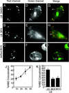
Evidence for a sorting pathway between TGN and phagosomes and its disruption by mycobacteria. Shown are live epifluorescent microscopy images of phagosomal acquisition of NBD-C6-sphingomyelin generated from NBD-C6-ceramide in the Golgi apparatus. (AC) Red fluorescence of latex beads or M. tuberculosis var. bovis bacillus Calmette–Guérin (BCG) expressing dsRed. LB, latex beads. (DF) Green fluorescence of NBD. (GI) Merged images of red and green fluorescence. After phagocytosis, cells were exposed to NBD-C6-ceramide for 30 min and incubated in the presence of defatted BSA to extract any NBD-sphingomyelin fluorescence from the plasma membrane. Cells were incubated in medium with DMSO solvent added (A, D, and G), in 5 μg/ml BFA (B, E, and H), or with medium alone (C, F, and I). (Size bar, 5 μm.) Filled arrowheads, examples of colocalization; open arrowheads, examples of no colocalization; asterisks, NBD-stained Golgi. (J) Time course of NBD-sphingomyelin acquisition by ingested latex beads (n = 1,495 phagosomes). The filled arrowheads exemplify phagosomes that colocalized with NBD-C6-sphingomyelin, and the open arrowheads exemplify phagosomes that did not colocalize with NBD-C6-sphingomyelin. (K) Quantitation of NBD-C6-sphingomyelin colocalization with phagosomes (n = 1,215 phagosomes) 4 h postinfection. The data are means ± SE of three separate experiments. ct, control; *, P < 0.001.
Figure 3
Figure 3
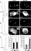
M. tuberculosis glycosylated phosphatidylinositol ManLAM inhibits delivery of syntaxin 6 and immature lysosomal hydrolase delivery to phagosomes. (A) Chemical structure of M. tuberculosis glycosylated phosphatidylinositol ManLAM. A, arabinose; M, mannose. The fatty acids are palmitoyl and tuberculostaroyl chains (34). (B) ManLAM precursor PIM. The arrows indicate several glycosylation steps adding the proximal mannose chain (M32), distal arabinose chain (A24), and several branched arabinose side chains capped with mannose (MxAy; M2). (C) A typical mammalian phosphatidylinositol (PtdIns) with the positions (–5) marked; these positions normally undergo phosphorylation in the process of directing intracellular trafficking. (D, H, F, and J) Green fluorescence of latex beads. (E and I) Syntaxin (Syn) 6 immunofluorescence. (G and K) Immature cathepsin D (iCat D) immunofluorescence. The arrowheads indicate phagosome colocalization with syntaxin 6 or immature cathepsin D. (Size bars, 5 μm.) Cells were allowed to phagocytize control beads (DG) or ManLAM-coated latex beads (HK). (L) Quantitation of syntaxin 6 and immature cathepsin D colocalization with control phagosomes (Con) or phagosomes coated with ManLAM 30 min postinfection (n = 1,376 phagosomes). (M) Quantitation of syntaxin 6 colocalization with control phagosomes (Con) or phagosomes containing latex beads coated with PIM, a ManLAM precursor (30 min postinfection; n = 547 phagosomes). The data are means ± SE from three separate experiments. *, P = 0.0125; **, P = 0.002.
Figure 4
Figure 4
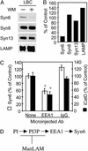
ManLAM interferes with the wortmannin-sensitive trafficking pathway from TGN to phagosomes. (A) Western blot analysis of purified LBC. Cultures were treated with 100 nM wortmannin (WM) 10 min after bead uptake by J774 cells and incubated with the inhibitor for 10 min before processing. LBC (5 μg of protein) were analyzed with antibodies against syntaxin (Syn) 6, syntaxin 8, syntaxin 13, and LAMP. (B) Quantitation of relative intensities of bands from a representative experiment shown in A (n = 3 experiments). (C) Microinjection of anti-EEA1 antibody reduces accumulation of syntaxin 6 and immature cathepsin D on latex bead phagosomes. Shown is quantitation of syntaxin 6 (n = 525 phagosomes) and immature cathepsin D (iCatD; n = 794 phagosomes) colocalization with latex bead phagosomes (30 min postinfection) in cells injected with anti-EEA1, control IgG, and uninjected control cells. The data are means ± SE of three separate experiments. *, P < 0.01. (D) Model of the inhibitory action of ManLAM. ManLAM disrupts the trafficking between the TGN and phagosomes. This phospholipid, structurally an analog of mammalian phosphatidylinositols (PI; see Fig. 3 AC), blocks the PI3P-dependent (PI3P) steps in trafficking.

References

    1. Meresse S, Steele-Mortimer O, Moreno E, Desjardins M, Finlay B, Gorvel J P. Nat Cell Biol. 1999;1:E183–E188. - PubMed
    1. Russell D G, Mwandumba H C, Rhoades E E. J Cell Biol. 2002;158:421–426. - PMC - PubMed
    1. Dye C, Scheele S, Dolin P, Pathania V, Raviglione M C. J Am Med Assoc. 1999;282:677–686. - PubMed
    1. Armstrong J A, Hart P D. J Exp Med. 1971;134:713–740. - PMC - PubMed
    1. Clemens D L, Horwitz M A. J Exp Med. 1995;181:257–270. - PMC - PubMed

Publication types

MeSH terms