. 2003 May;71(5):2563-70.
doi: 10.1128/IAI.71.5.2563-2570.2003.

Cleavage of the human immunoglobulin A1 (IgA1) hinge region by IgA1 proteases requires structures in the Fc region of IgA

Affiliations

Cleavage of the human immunoglobulin A1 (IgA1) hinge region by IgA1 proteases requires structures in the Fc region of IgA

Koteswara R Chintalacharuvu et al. Infect Immun. 2003 May.

Abstract

Secretory immunoglobulin A (IgA) protects the mucosal surfaces against inhaled and ingested pathogens. Many pathogenic bacteria produce IgA1 proteases that cleave in the hinge of IgA1, thus separating the Fab region from the Fc region and making IgA ineffective. Here, we show that Haemophilus influenzae type 1 and Neisseria gonorrhoeae type 2 IgA1 proteases cleave the IgA1 hinge in the context of the constant region of IgA1 or IgA2m(1) but not in the context of IgG2. Both C(alpha)2 and C(alpha)3 but not C(alpha)1 are required for the cleavage of the IgA1 hinge by H. influenzae and N. gonorrhoeae proteases. While there was no difference in the cleavage kinetics between wild-type IgA1 and IgA1 containing only the first GalNAc residue of the O-linked glycans, the absence of N-linked glycans in the Fc increased the ability of the N. gonorrhoeae protease to cleave the IgA1 hinge. Taken together, these results suggest that, in addition to the IgA1 hinge, structures in the Fc region of IgA are required for the recognition and cleavage of IgA1 by the H. influenzae and N. gonorrhoeae proteases.


Figures
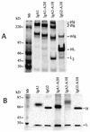
FIG. 1.
FIG. 1.
SDS-PAGE analysis of secreted wild-type and hinge exchanged antibodies. Transfectants were biosynthetically labeled with [35S]methionine for 16 h, and the labeled Igs were precipitated from the culture supernatants with dansylated BSA coupled to Sepharose. Labeled proteins were analyzed by SDS-PAGE in 5% PO4 gels under nonreducing conditions (A) and on 12.5% Tris-glycine gels after reducing with β-mercaptoethanol (B). pIg, Igs larger than dimers; dIg, dimeric Igs; mIg, monomeric Igs; Mr, molecular weight marker.
FIG. 2.
FIG. 2.
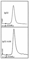
Fast protein liquid chromatography analysis of wild-type IgG2 (A) and IgG2-A1H (B). A total of 100 μg of purified protein was analyzed by gel filtration on a 30- by 1.5-cm Superose 6 column in PBS. Note that both proteins migrated as a single peak with a retention volume of 17.5 ml.
FIG. 3.
FIG. 3.
SDS-PAGE analysis of wild-type IgG2, IgA1, and IgA1-A2H and the IgA2-A1H and IgG2-A1H proteins digested with H. influenzae (HI) and N. gonorrhoeae (NG) IgA1 proteases. 35[S]Methionine-labeled proteins were mixed with 2 μg of unlabeled IgA1-2 μl of concentrated protease supernatant in 50 mM Tris buffer, pH 7.5, containing 0.05% NaN3 and 10 mM EDTA in a total volume of 20 μl and incubated at 37°C for 2 h. The samples were boiled in sample buffer containing β-mercaptoethanol and analyzed by SDS-PAGE in 12.5% Tris-glycine gels. Mr, molecular weight marker.
FIG. 4.
FIG. 4.
SDS-PAGE analysis of domain-exchanged proteins digested with H. influenzae (HI) and N. gonorrhoeae (NG) IgA1 proteases. (A through C) a mixture containing 35[S]methionine-labeled proteins, 2 μg of unlabeled IgA1, and 2 μl of protease for each time point was incubated in a 37°C water bath. At various time points, aliquots were removed and the proteolysis was stopped and analyzed by SDS-PAGE as described in the legend to Fig. 3. (D) The H-chain and L-chain expression vectors were expressed in 293T cells and biosynthetically labeled with [35S]methionine as described in Materials and Methods. Labeled protein was digested and analyzed as described in the legend to Fig. 3. To eliminate the possibility of differences due to expression in different cell types, the wild-type IgA1 H chain and L chain were expressed in 293T cells and biosynthetically labeled and the labeled protein was digested with proteases and analyzed as described in the legend to Fig. 3. (data not shown).
FIG. 5.
FIG. 5.
Kinetics of digestion of wild-type IgA1 and IgA1 lacking N-linked glycans digested with H. influenzae (HI) (A) and N. gonorrhoeae (NG) (B) proteases. 35[S]Methionine-labeled IgA1 lacking N-linked glycans was digested and analyzed by SDS-PAGE as described in the legend to Fig. 3. Molecular weights are indicated to the left of the gels.
FIG. 6.
FIG. 6.
Degradation of wild-type IgA1, IgA1 lacking N-linked glycans, and IgA1 lacking N-linked glycans and containing truncated O-linked glycans by H. influenzae (A) and N. gonorrhoeae (B) proteases. Bands in Fig. 5 were quantified by spot densitometry. Since the H. influenzae and N. gonorrhoeae IgA1 proteases do not cleave L chains, intact H-chain values were normalized against the L-chain values obtained in order to control for loading differences from lane to lane. wt, wild type.

References

    1. Aalberse, R. C., and J. Schuurman. 2002. IgG4 breaks the rules. Immunology 105:9-19. - PMC - PubMed
    1. Baenziger, J., and S. Kornfeld. 1974. Structure of the carbohydrate units of IgA1 immunoglobulin. I. Composition, glycopeptide isolation, and structure of the asparagine-linked oligosaccharide units. J. Biol. Chem. 249:7260-7269. - PubMed
    1. Baenziger, J., and S. Kornfeld. 1974. Structure of the carbohydrate units of IgA1 immunoglobulin. II. Structure of the O-glycosidically linked oligosaccharide units. J. Biol. Chem. 249:7270-7281. - PubMed
    1. Chintalacharuvu, K. R., M. Raines, and S. L. Morrison. 1994. Divergence of human alpha-chain constant region gene sequences. A novel recombinant alpha 2 gene. J. Immunol. 152:5299-5304. - PubMed
    1. Chintalacharuvu, K. R., L. J. Yu, N. Bhola, K. Kobayashi, C. Z. Fernandez, and S. L. Morrison. 2002. Cysteine residues required for the attachment of the light chain in human IgA2. J. Immunol. 169:5072-5077. - PubMed

Publication types

MeSH terms

LinkOut - more resources