. 2003 May;71(5):2634-42.
doi: 10.1128/IAI.71.5.2634-2642.2003.

CC chemokines mediate leukocyte trafficking into the central nervous system during murine neurocysticercosis: role of gamma delta T cells in amplification of the host immune response

Affiliations

CC chemokines mediate leukocyte trafficking into the central nervous system during murine neurocysticercosis: role of gamma delta T cells in amplification of the host immune response

Astrid E Cardona et al. Infect Immun. 2003 May.

Abstract

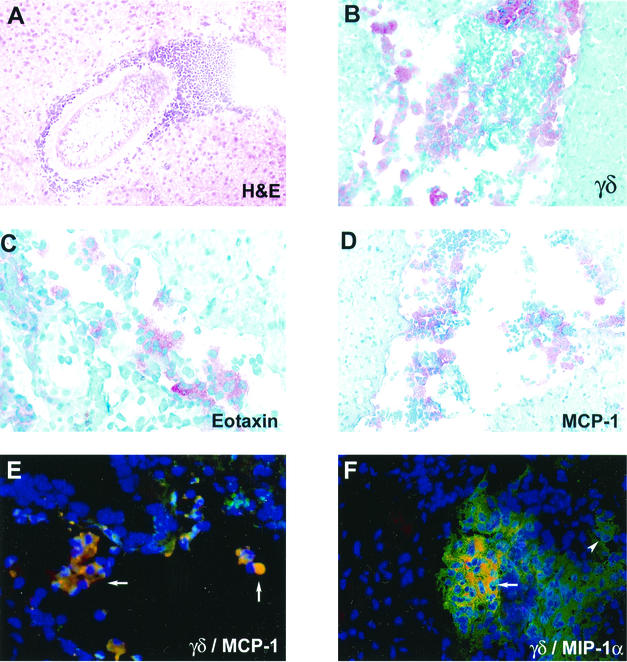
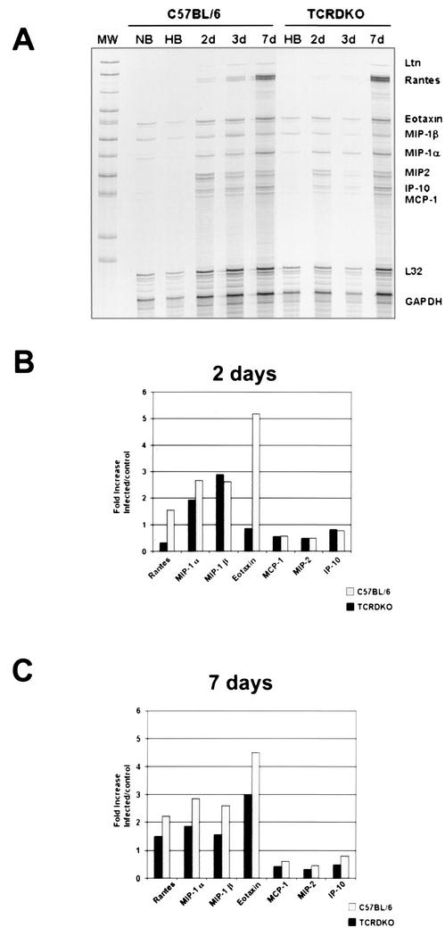
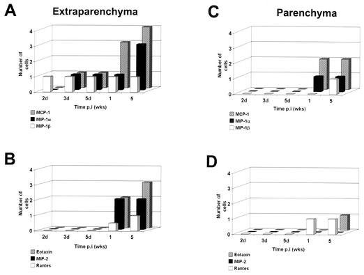
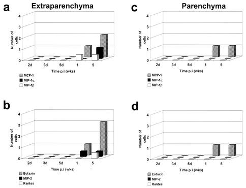
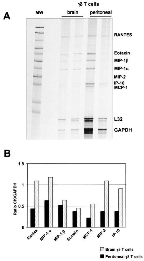
According to a previous report, the degree of the host immune response highly correlates with severity of the disease in the murine model for neurocysticercosis. In wild-type mice, Mesocestoides corti infection induced a rapid and extensive accumulation of gamma delta T cells and macrophages in the brain. NK cells, dendritic cells, alpha beta T cells, and B cells were also recruited to the brain but at lower levels. In contrast, gamma delta T-cell-deficient mice exhibited decreased cellular infiltration and reduced central nervous system (CNS) pathology. To understand the mechanisms of leukocyte recruitment into the CNS, chemokine expression was analyzed in infected brains in the present study. MCP-1 (CCL2), MIP-1 alpha (CCL3), and MIP-1 beta (CCL4) were up-regulated within 2 days after M. corti infection. Protein expression of RANTES (CCL5), eotaxin (CCL11), and MIP-2 was detected later, at 1 week postinfection. Correlating with the decreased cellular infiltration, delta chain T-cell receptor-deficient (TCR delta(-/-)) mice exhibited substantially reduced levels of most of the chemokines analyzed (with the exception of eotaxin). The results suggest that gamma delta T cells play an important role in the CNS immune response by producing chemokines such as MCP-1 and MIP-1 alpha, enhancing leukocyte trafficking into the brain during murine neurocysticercosis.


Figures

FIG. 1.
FIG. 1.
Cellular infiltration and chemokine expression in the CNS during murine NCC. (A) Hematoxylin and eosin (H&E)-stained brain section from C57BL/6 mouse at 3 weeks p.i. (B) γδ T cells from C57BL/6 brain infected for 1 week as detected by immunohistochemistry. (C and D) Eotaxin (C)- and MCP-1 (D)-expressing cells from C57BL/6 brain cryosections from infected mice on day 3 as detected by immunocytochemistry. (E and F) Double-immunofluorescence assays were performed on brain cryosections of wild-type mice at 1 week p.i.; merged pictures are shown. MCP-1 (E) and MIP1-α (F) producing γδ T cells in a ventricular infiltrate are shown in yellow as double positive (arrows). γδ T cells (single positive) were detected with the GL3-phycoerythrin-conjugated antibody (red fluorescence), and chemokine-producing cells (single positive) were detected with a green fluorescent conjugate. The arrowhead points to single-positive cells. Cell nuclei visualized in blue (DAPI staining) are shown.
FIG. 2.
FIG. 2.
Predominance of CC chemokines in the brain after M. corti infection as assayed by RPA. Total RNA obtained from brain lymphocytes was used to detect the expression of several chemokines. (A) Autoradiograph of the RPA using the mCK5 template from Pharmingen at 2, 3, and 7 days (2d, 3d, and 7d, respectively) p.i. Normal brains (NB) and HBSS-inoculated brains (HB) were used as controls. MW, molecular weight markers. (B) Densitometry analysis of the chemokine expression at 2 days p.i. Samples were normalized to the GAPDH gene, and the infected sample cell/HBSS control cell ratios are shown. (C) Densitometry analysis at 7 days p.i. Data represent the values calculated for four mice at each time point.
FIG. 3.
FIG. 3.
Chemokine response in the brains of C57BL/6 mice. Mouse brains obtained after intracranial inoculation with M. corti metacestodes were analyzed by immunohistochemistry for the presence of several chemokines. (A and B) Positive cells in extraparenchymal infiltrates (subarachnoid spaces, meninges, and ventricles) were counted, and the number of positive cells was scored as follows: 1, 1 to 100 cells; 2, 100 to 300 cells; and 3, 300 to 500 cells. (C and D) Chemokine-producing cells associated with parenchymal infiltrates. The results indicate the average of results for at least two mice at each time point.
FIG. 4.
FIG. 4.
Chemokine response in the brain of γδ T-cell-deficient mice (TCRδ−/−) mice. Two mouse brains obtained after intracranial inoculation with M. corti metacestodes were analyzed by immunohistochemistry for the presence of several chemokines. (a and b) Positive cells in extra-parenchymal infiltrates (including subarachnoid spaces, meninges, and ventricles) were counted, and the number of positive cells was scored as follows: 1, 1 to 100 cells; 2, 100 to 300 cells; 3, 300 to 500 cells. (c and d) Chemokine-producing cells associated with parenchymal infiltrates.
FIG. 5.
FIG. 5.
Chemokine analysis in isolated γδ T cells. Using an RPA, brain and peritoneal γδ T cells were analyzed for the expression of several chemokines. (A) Autoradiograph of total RNA from γδ T lymphocytes. MW, molecular weight markers. (B) Densitometry analysis of chemokine (CK) expression. Samples were normalized to the GAPDH to indicate relative mRNA abundance.

References

    1. Asensio, V. C., and I. L. Campbell. 1997. Chemokine gene expression in the brains of mice with lymphocytic choriomeningitis. J. Virol. 71:7832-7840. - PMC - PubMed
    1. Asensio, V. C., and I. L. Campbell. 1999. Chemokines in the CNS: plurifunctional mediators in diverse states. Trends Neurosci. 22:1453-1458. - PubMed
    1. Asensio, V. C., C. Kincaid, and I. L. Campbell. 1999. Chemokines and the inflammatory response to viral infection in the central nervous system with a focus on lymphocytic choriomeningitis virus. J. Neurovirol. 5:65-75. - PubMed
    1. Azuara, V., J. P. Levraud, M. P. Lembezat, and P. Pereira. 1997. A novel subset of adult γδ thymocytes that secretes a distinct pattern of cytokines and expresses a very restricted T cell repertoire. Eur. J. Immunol. 27:544-553. - PubMed
    1. Baggiolini, M. 1998. Chemokines and leukocyte traffic. Nature 392:565-568. - PubMed

Publication types

LinkOut - more resources