. 2003 May;71(5):2684-92.
doi: 10.1128/IAI.71.5.2684-2692.2003.

Persistence of mucosal mast cells and eosinophils in Shigella-infected children

Affiliations

Persistence of mucosal mast cells and eosinophils in Shigella-infected children

Rubhana Raqib et al. Infect Immun. 2003 May.

Abstract

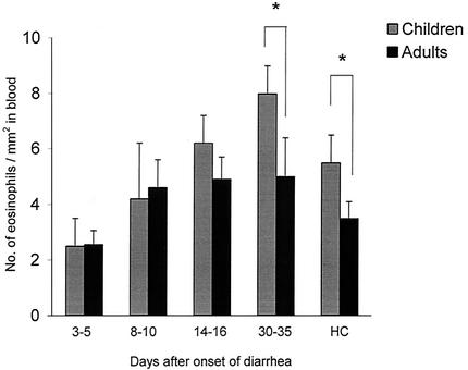
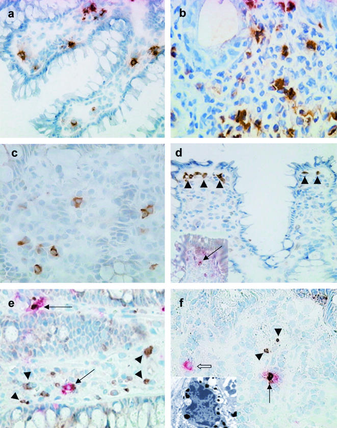
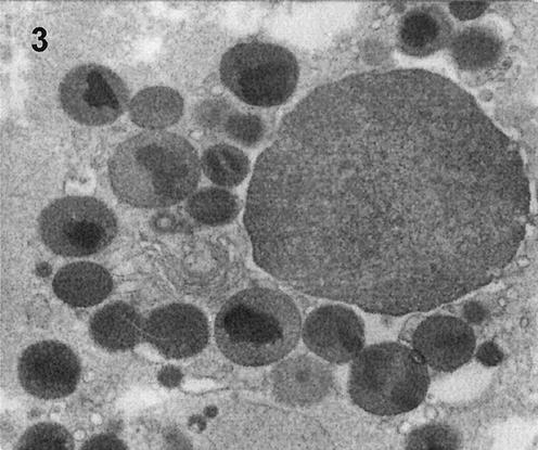
Cells of the innate immune system and their mediators were studied at the single-cell level in the rectums of pediatric and adult patients with Shigella infection to better understand why children are at higher risk for severe infection. Adult patients had increased infiltration of mucosal mast cells (MMC) at the acute stage (3 to 5 days after the onset of diarrhea) and eosinophils in early convalescence (14 to 16 days after onset). Increased expression of stem cell factor and prostaglandin H synthase-1 (PGHS-1) was associated with increased tryptase-K(i)67-double-positive MMC in the acute stage and increased apoptosis of MMC, which led to a rapid decline in early convalescence. The eosinophils demonstrated increased expression of major basic protein (MBP), eotaxin, and CCR3, as well as increased necrotic death. The neutrophils showed enhanced alpha-defensin and lactoferrin expression in the acute phase. In contrast to adults, the pediatric patients demonstrated delayed accumulation of mast cells and eosinophils, while alpha-defensin expression persisted during convalescence. In contrast, neutrophil counts and lactoferrin expression were reduced in children compared to adults. The results suggest that children with shigellosis have a persistent activation of the innate immune response in the convalescent phase, indicating delayed elimination of Shigella antigens compared to adults.


Figures

FIG. 1.
FIG. 1.
Comparison of eosinophil counts in blood in pediatric and adult patients with shigellosis at various times after onset of diarrhea. The eosinophil counts in children (shaded bar) and in adults (solid bar) are given as geometric means with standard errors. Blood samples were collected 3 to 5, 8 to 10, 14 to 16, and 33 to 35 days after the onset of diarrhea. HC, healthy controls. The asterisks indicate a significant difference between adult and pediatric patients (P < 0.05).
FIG.2.
FIG.2.
Rectal biopsy sections from adult and pediatric patients showing localization of eosinophils, mast cells, and various mediators, as well as proliferating and apoptotic mast cells, during the course of Shigella infection. (a) Eosinophils expressing MBP were significantly increased in the rectum of a Shigella-infected pediatric patient during late convalescence (30 to 35 days after the onset of disease). Magnification, ×380. (b) Eotaxin expression was markedly upregulated during acute Shigella infection (3 to 5 days after the onset of diarrhea) in a pediatric patient. Eotaxin expression was localized to the eosinophils, as well as neutrophils, as is evident from the morphological appearance. Magnification, ×855. (c) Immunohistochemical staining revealed increased expression of CCR3 in rectal tissue during acute Shigella infection (3 to 5 days after the onset of disease) in an adult. The staining was localized primarily to the cytoplasm of the cells. Magnification, ×570. (d) Immunohistochemical staining delineates cells underlying the surface epithelium expressing PGHS-1 during acute dysentery (3 to 5 days after the onset of disease) in an adult patient. The positive cells were in close proximity to each other (arrowheads). The inset shows a chymase-positive mast cell (deep pink) expressing PGHS-1 (brown) just below the surface epithelium, as shown by double staining (arrow). Magnification, ×380. (e) During the early stage of acute Shigella infection (3 to 5 days after the onset of diarrhea), increased numbers of Ki67-positive cells (brown; arrowheads) were present in the lamina propria and in the crypt epithelial lining in a pediatric patient. Chymase-positive mast cells (deep pink) also stained for anti-Ki67 antibody (brown), indicating active proliferation of mast cells (arrows). Magnification, ×570. (f) During acute shigellosis (3 to 5 days after the onset of diarrhea), apoptotic (TUNEL-positive [brown; arrowheads]) cells were seen in the lamina propria in an adult patient. Double immunohistochemical staining showed an apoptotic (brown) chymase-positive mast cell (deep pink, thin arrow) in the proximity of an unaffected mast cell (deep pink; open arrow) in the lamina propria. Magnification, ×380. In the inset, ultrastructural analysis reveals an apoptotic mast cell. The mast cell shows nuclear pyknosis, without cytoplasmic-organelle swelling, suggestive of early apoptosis. Magnification, ×7,980.
FIG. 3.
FIG. 3.
Electron micrograph of a necrotic eosinophil in the lamina propria in the rectal mucosa during acute Shigella infection (3 to 5 days after the onset of diarrhea) in a pediatric patient. Magnification, ×19,000.

References

    1. Abraham, S. N., and R. Malaviya. 2000. Mast cell modulation of the innate immune response to enterobacterial infection. Adv. Exp. Med. Biol. 479:91-105. - PubMed
    1. Abraham, S. N., and R. Malaviya. 1997. Mast cells in infection and immunity. Infect. Immun. 65:3501-3508. - PMC - PubMed
    1. Anand, B. S., V. Malhotra, S. K. Bhattacharya, P. Datta, D. Datta, D. Sen, M. K. Bhattacharya, P. P. Mukherjee, and S. C. Pal. 1986. Rectal histology in acute bacillary dysentery. Gastroenterology 90:654-660. - PubMed
    1. Artis, D., N. E. Humphreys, C. S. Potten, N. Wagner, W. Muller, J. R. McDermott, R. K. Grencis, and K. J. Else. 2000. Beta7 integrin-deficient mice: delayed leukocyte recruitment and attenuated protective immunity in the small intestine during enteric helminth infection. Eur. J. Immunol. 30:1656-1664. - PubMed
    1. Chertov, O., D. Yang, O. M. Howard, and J. J. Oppenheim. 2000. Leukocyte granule proteins mobilize innate host defenses and adaptive immune responses. Immunol. Rev. 177:68-78. - PubMed

Publication types

LinkOut - more resources