Comparative Study
. 2003 May;4(5):517-22.
doi: 10.1038/sj.embor.embor824.

Otubains: a new family of cysteine proteases in the ubiquitin pathway

Affiliations
Comparative Study

Otubains: a new family of cysteine proteases in the ubiquitin pathway

Maxim Y Balakirev et al. EMBO Rep. 2003 May.

Abstract

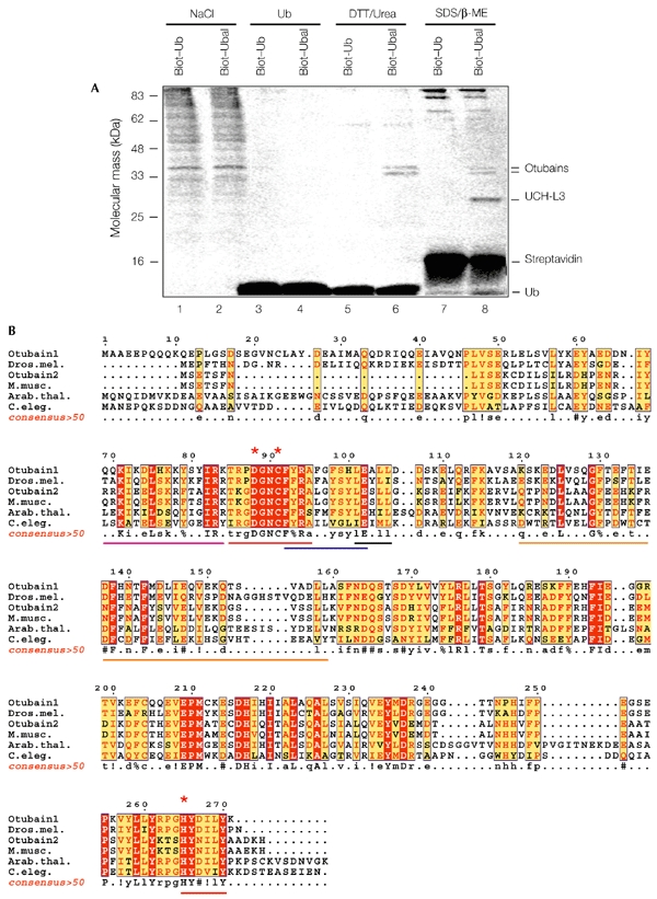
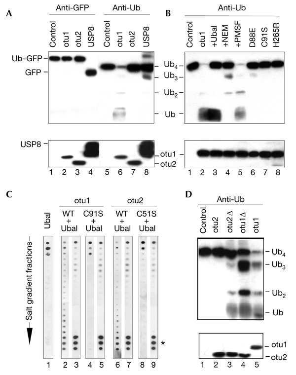
The modification of cellular proteins by ubiquitin (Ub) is an important event that underlies protein stability and function in eukaryotes. Protein ubiquitylation is a dynamic and reversible process; attached Ub can be removed by deubiquitylating enzymes (DUBs), a heterogeneous group of cysteine proteases that cleave proteins precisely at the Ub-protein bond. Two families of DUBs have been identified previously. Here, we describe new, highly specific Ub iso-peptidases, that have no sequence homology to known DUBs, but which belong to the OTU (ovarian tumour) superfamily of proteins. Two novel proteins were isolated from HeLa cells by affinity purification using the DUB-specific inhibitor, Ub aldehyde (Ubal). We have named these proteins otubain 1 and otubain 2, for OTU-domain Ubal-binding protein. Functional analysis of otubains shows that the OTU domain contains an active cysteine protease site.


Figures

Figure 1
Figure 1
Identification of novel ubiquitin-aldehyde-binding proteins. (A) Ubiqutin aldehyde (Ubal)-affinity purification of otubains. Cell lysates were incubated with biotin–ubiquitin (biot–Ub) or biot–Ubal (20 ng mg−1 of protein; for 1 h at 4 °C), and loaded onto streptavidin–agarose columns. After extensive washing, the bound proteins were eluted with 1.5 M NaCl in suspension buffer (SB; lanes 1 and 2), 1 mg ml−1 Ub in SB (lanes 3 and 4), 100 mM dithiothreitol (DTT) and 4 M urea in SB (lanes 5 and 6), or 1 × Laemmli buffer (lanes 7 and 8). The eluted proteins were resolved by SDS–polyacrylamide gel electrophoresis and stained with Coomassie blue. The proteins identified by mass spectrometry are indicated. (B) Clustal W sequence alignment of the otubain-family proteins, edited with ESPript software (Gouet et al., 1999). The predicted proteins are from humans (GenBank accession numbers: for otubain 1; for otubain 2), Drosophila melanogaster (Dros. mel.; ), Mus musculus (M. musc.; ), Arabidopsis thaliana (Arab. thal.; ) and Caenorhabditis elegans (C. eleg.; ). The red asterisks indicate the putative catalytic triad of the cysteine protease, and the lines below the sequences mark the OTU (ovarian tumour) domain (red), putative nuclear localization signal (magenta), Ub interaction motif (UIM)-like motif Φ-xx-A-xxxs-xx-Ac (where Φ indicates an aromatic amino acid, x indicates any amino acid and Ac indicates an acidic amino acid; blue), Ub-associated (UBA)-like domain (orange) and the LxxLL motif (black). β-ME, β-mercaptoethanol. A consensus >50 sequence (a sequence showing residues that are more than 50% conserved) was generated using ESPript software. Uppercase letters in the consensus>50 sequence and red shading in the other sequences indicate identity; lowercase letters in the consensus >50 sequence and yellow shading in the other sequences indicate a consensus level of >50%; ! indicates I or V; % indicates F or Y; # indicates N, D, Q or E.
Figure 2
Figure 2
Otubains are deubiquitylating enzymes. (A) Proteolysis of ubiquitin–green fluorescent protein (Ub–GFP; lanes 1–4) and tetra-Ub (lanes 5–8) by otubains (otu1 and otu2) and ubiquitin-specific protease 8 (USP8). Assays were carried out at 37 °C for 30 min, in 20 μl of assay buffer containing the protein substrate (0.1–1.0 μM) and a protease (10–100 nM). Substrate cleavage was analysed by western blotting with anti-GFP (lanes 1–4) and anti-Ub (lanes 5–8) antibodies. The lower blot shows the proteases in each reaction probed with anti-His6 (for the otubains) and anti-glutathione-S-transferase (anti-GST; for GST–USP8) antibodies. (B) Protease inhibition profile of otubain 1. Effect of different inhibitors (lanes 1–5) and OTU (ovarian tumour)-domain mutations (lanes 6–8) on the processing of tetra-Ub by otubain 1. Reaction conditions were the same as in Fig. 2A, except that 0.5 μM Ub aldehyde (Ubal; lane 3), 0.5 μM NEM (N-ethylmaleimide; lane 4), or 1 mM PMSF (phenylmethylsulphonyl fluoride; lane 5) were added, as indicated. (C) Otubain–Ubal interaction. Wild-type otubains (otu1 and otu2: WT) and their activesite mutants (otu1 and otu2: C91/51S) were incubated with Ubal, and the reaction mixtures were separated by fast performance liquid chromatography, using an anion-exchange column. The fractions were eluted over a salt gradient and dot-blotted and probed in parallel, using the anti-Ub antibody (lanes 1, 2, 4, 6 and 8) and affinity-purified anti-otubain antibodies (lanes 3, 5, 7 and 9). Ubal alone was eluted at the beginning of the gradient (lane 1). The formation of otubain–Ubal complexes was seen by co-purification of Ubal with otubains (asterisk, lanes 2, 3, 6 and 7). Complex formation did not change the elution profile of the otubains (not shown). The active-site mutants of otubains did not interact with Ubal (lanes 4, 5, 8 and 9). (D) Effect of otubain truncations on tetra-Ub proteolysis. Reaction conditions were the same as in Fig. 2A. The lower panel shows the otubains blotted with the anti-His6 antibody. Otubain 1Δ (otu1Δ) shows slightly higher SDS–polyacrylamide gel electrophoreis mobility than otu 2 and otubain 2Δ1–229 (otu2Δ).
Figure 3
Figure 3
Expression of otubains in tissues and cultured cells. (A) RT–PCR (polymerase chain reaction with reverse transcription) analysis of otubains, performed on a panel of complementary DNAs from various human tissues (OriGene). Note the two forms of the otubain 1 (otu1) transcript from leukocytes, and that the highest level of otubain 2 (otu2) expression was in brain tissue. (B) Western blot analysis of otubains in human tissues (Protein Medley; Clontech) using affinity-purified anti-otubain antibodies. (C) Transfection of HeLa cells with wild-type otubains (otu1 and otu2: WT), their active-site mutants (otu1 and otu2: C91/51S), and antisense cDNAs (otu1 and otu2: AS) on intracellular ubiquitin (Ub)–protein conjugates. Note the slight decrease in Ub-conjugates in otubain 1-transfected cells (otu1, WT lane) and the disappearance of endogenous otubain 1 induced by antisense cDNA (otu1; AS; middle blot). Tub, tubulin; uH2A, monoubiquitylated histone 2A.
Figure 4
Figure 4
Role of the otubain-like domain of the A20 protein in the regulation of tumour necrosis factor signalling. (A) The TRAF (TNF-receptor-associated factor)-binding domain of A20 homologues (cellular zinc-finger anti-nuclear factor-κB (NF-κB) protein (Cezanne) and TRAF-binding-domain protein (TRABID); Evans et al., 2001) is shown, aligned with the otubain activesite (represented as a consensus>50 sequence (a sequence showing residues that are more than 50% conserved) as shown in Fig. 1B). The activesite Asp in the TRABID protein is replaced by an Ala residue. Uppercase letters in the consensus>50 sequences and red shading in the other sequences indicate identity; lowercase letters in the consensus >50 sequences and yellow shading indicate a consensus level of >50%; # indicates N, D, Q or E; red asterisks indicate the putative catalytic triad of the cysteine protease. (B) Effect of mutations in active-site cysteine residues on the NF-κB-inhibitory function of the A20 protein. A reporter-gene assay was performed using the firefly luciferase (Fluc) construct κB3–luc and the Renilla luciferase (Rluc) construct pRL–TK. HeLa cells were co-transfected with 100 ng of κB3–luc, 100 ng of pRL-TK, and the indicated amounts (in nanograms) of the haemagglutinin (HA)–A20 and HA–A20(C103S) constructs. The total amount of transfected DNA was kept constant at 400 ng by adding pHM6 empty vector. NF-κB was induced 18 h after transfection by stimulation with tumour necrosis factor (TNF)-α for 6 h, and its activity is represented as normalized Fluc/Rluc activity. The result shown is an average of two experiments. The blot above the graph shows the expression of A20 proteins in transfected cells as detected by the anti-HA antibody.

References

    1. Balakirev M., Jaquinod M., Haas A. & Chroboczek J. (2002) Deubiquitinating function of adenovirus proteinase. J. Virol., 76, 6323–6331. - PMC - PubMed
    1. Beyaert R., Heyninck K. & Van Huffel S. (2000) A20 and A20-binding proteins as cellular inhibitors of nuclear factor-κB-dependent gene expression and apoptosis. Biochem. Pharmacol., 60, 1143–1151. - PubMed
    1. Borodovsky A., Ovaa H., Kolli N., Gan-Erdene T., Wilkinson K.D., Ploegh H.L. & Kessler B.M. (2002) Chemistry-based functional proteomics reveals novel members of the deubiquitinating enzyme family. Chem. Biol., 9, 1149–1159. - PubMed
    1. Chen X., Zhang B. & Fischer J.A. (2002) A specific protein substrate for a deubiquitinating enzyme: Liquid facets is the substrate of Fat facets. Genes Dev., 16, 289–294. - PMC - PubMed
    1. D'Andrea A. & Pellman D. (1998) Deubiquitinating enzymes: a new class of biological regulators. Crit. Rev. Biochem. Mol. Biol., 33, 337–352. - PubMed

Publication types

MeSH terms