

Induction of caspase-dependent apoptosis by betanodaviruses GGNNV and demonstration of protein alpha as an apoptosis inducer
- PMID: 12706091
- PMCID: PMC7126978
- DOI: 10.1016/s0042-6822(02)00098-3
Induction of caspase-dependent apoptosis by betanodaviruses GGNNV and demonstration of protein alpha as an apoptosis inducer
Abstract
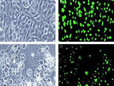
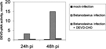
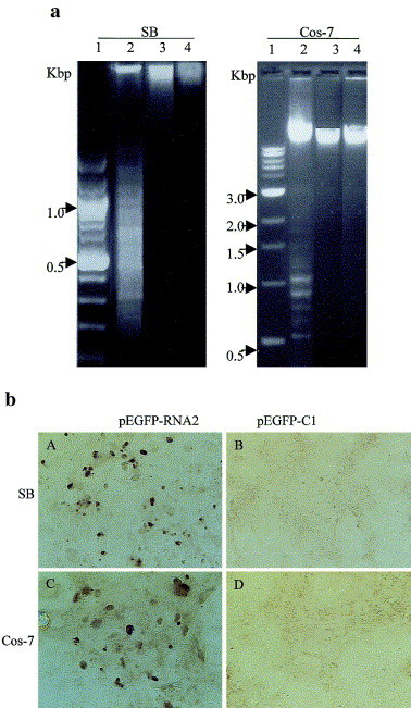
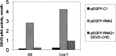
Betanodaviruses, members of the Nodaviridae family, are the causative agents of viral nervous necrosis in fish and infection by which cause high mortality in larvae and juveniles in a wide range of marine fish species in Asia, Europe, Australia, Martinique, and Tahit. Greasy grouper (Epinephelus tauvina) nervous necrosis viruses (GGNNV) were investigated for their apoptotic activity in culture cells. GGNNV infection of sea bass (SB) cells appeared to induce a typical cytopathic effect (CPE), i.e., cytoplasmic vacuolation, thinning, rounding up, detachment of infected cells from the cultured dish, and eventually cell lysis and death. The infected SB cells underwent DNA fragmentation and stained positive in terminal deoxynucleotidyl transferase (TdT)-mediated dUTP nick-end labeling (TUNEL) assay, suggesting that GGNNV infection induced apoptosis in SB cells. In addition, GGNNV-infected SB cells showed an increased activity of caspase-8-like proteases (IETDase) and caspase-3-like proteases (IETDase), whereas inhibitor of caspase-8 and caspase-3 reduced GGNNV-induced apoptosis. This suggests that GGNNV may promote apoptosis via the extrinsic pathway in SB cells. Protein alpha, the precursor of GGNNV capsid proteins, was transiently expressed in SB and Cos-7 cells. The DNA fragmentation and TUNEL positive signal were apparent in SB and Cos-7 cells expressing protein alpha, suggesting that protein alpha may serve as an apoptotic inducer in these cells. Moreover, expression of protein alpha resulted in the activation of caspase-3-like proteases in both cells, which could be inhibited by a caspase-3-like protease specific inhibitor DEVD-CHO peptide. These results suggest that fish caspases are important elements in GGNNV-meditated apoptosis.
Figures


Similar articles
-
Membrane association of greasy grouper nervous necrosis virus protein A and characterization of its mitochondrial localization targeting signal.J Virol. 2004 Jun;78(12):6498-508. doi: 10.1128/JVI.78.12.6498-6508.2004. J Virol. 2004. PMID: 15163743 Free PMC article.
-
Identification of nucleolus localization signal of betanodavirus GGNNV protein alpha.Virology. 2003 Feb 15;306(2):225-35. doi: 10.1016/s0042-6822(02)00081-8. Virology. 2003. PMID: 12642096 Free PMC article.
-
GSIV serine/threonine kinase can induce apoptotic cell death via p53 and pro-apoptotic gene Bax upregulation in fish cells.Apoptosis. 2016 Apr;21(4):443-58. doi: 10.1007/s10495-016-1219-4. Apoptosis. 2016. PMID: 26833308
-
Apoptosis during ectromelia orthopoxvirus infection is DEVDase dependent: in vitro and in vivo studies.Microbes Infect. 2002 May;4(6):599-611. doi: 10.1016/s1286-4579(02)01578-2. Microbes Infect. 2002. PMID: 12048029
-
Characterization of virus-like particles assembled in a recombinant baculovirus system expressing the capsid protein of a fish nodavirus.Virology. 2001 Nov 10;290(1):50-8. doi: 10.1006/viro.2001.1157. Virology. 2001. PMID: 11883005
Cited by
-
Cloning of crystallin from orange-spotted grouper and characterization of its activity as potential protective agent.Results Immunol. 2011 Sep 9;1(1):60-9. doi: 10.1016/j.rinim.2011.08.005. eCollection 2011. Results Immunol. 2011. PMID: 24371554 Free PMC article.
-
Betanodavirus: Mitochondrial disruption and necrotic cell death.World J Virol. 2013 Feb 12;2(1):1-5. doi: 10.5501/wjv.v2.i1.1. World J Virol. 2013. PMID: 24175224 Free PMC article.
-
Susceptibility of Chinese Perch Brain (CPB) Cell and Mandarin Fish to Red-Spotted Grouper Nervous Necrosis Virus (RGNNV) Infection.Int J Mol Sci. 2016 May 19;17(5):740. doi: 10.3390/ijms17050740. Int J Mol Sci. 2016. PMID: 27213348 Free PMC article.
-
Betanodavirus B2 is an RNA interference antagonist that facilitates intracellular viral RNA accumulation.J Virol. 2006 Jan;80(1):85-94. doi: 10.1128/JVI.80.1.85-94.2006. J Virol. 2006. PMID: 16352533 Free PMC article.
-
Betanodavirus B2 causes ATP depletion-induced cell death via mitochondrial targeting and complex II inhibition in vitro and in vivo.J Biol Chem. 2010 Dec 17;285(51):39801-10. doi: 10.1074/jbc.M110.164988. Epub 2010 Sep 24. J Biol Chem. 2010. PMID: 20870718 Free PMC article.
References
-
- Adams J.M., Cory S. The Bcl-2 protein family: arbiters of cell survival. Science. 1998;281:1322–1326. - PubMed
-
- Agol V.I., Belov G.A., Bienz K., Egger D., Kolesnikova M.S., Raikhlin N.T., Romanova L.I., Smirnova E.A., Tolskaya E.A. Two types of death of poliovirus-infected cells: caspase involvement in the apoptosis but not cytopathic effect. Virology. 1998;252:343–353. - PubMed
-
- Ball L.A., Hendry D.A., Johnson J.E., Rueckert R.R., Scotti P.D. Family Nodaviridae. In: van Regenmortel M.H.V., Fauquet C.M., Bishop D.H.L., Carstens E.B., Estes M.K., Lemon S.M., Maniloff J., Mayo M.A., McGeoch D.J., Pringle C.R., Wickner R.B., editors. Virus Taxonomy. Seventh Report of the International Committee for the Taxonomy of Viruses. Academic Press; San Diego: 2000. pp. 747–755.
-
- Chong S.Y., Ngoh G.H., Ng M.K., Chu K.T. Growth of lymphocystis virus in a sea bass, Lates calcarifer (Bloch) cell line. Singapore Vet. J. 1987;11:78–85.
Publication types
MeSH terms
Substances
LinkOut - more resources
Full Text Sources
Research Materials
Miscellaneous
