. 2003 May;162(5):1503-13.
doi: 10.1016/S0002-9440(10)64283-6.

Aberrant expression and activation of the thrombin receptor protease-activated receptor-1 induces cell proliferation and motility in human colon cancer cells

Affiliations

Aberrant expression and activation of the thrombin receptor protease-activated receptor-1 induces cell proliferation and motility in human colon cancer cells

Dalila Darmoul et al. Am J Pathol. 2003 May.

Abstract

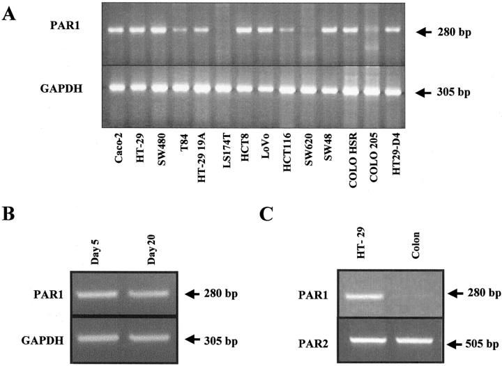
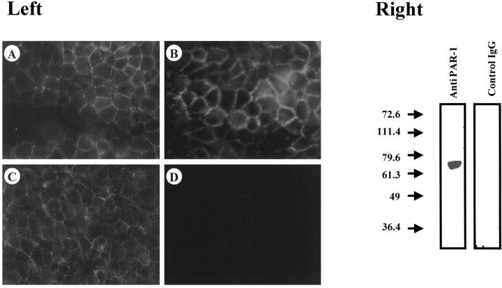
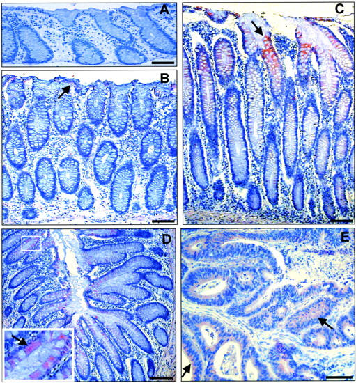
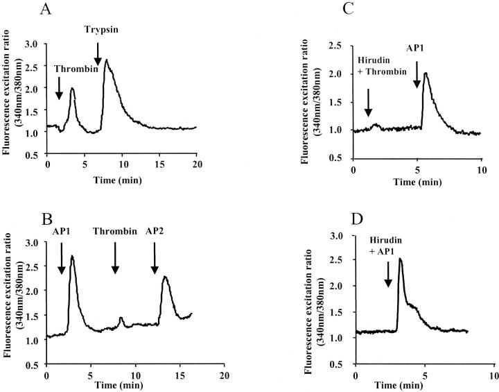
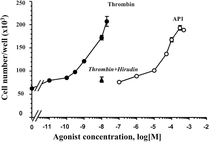
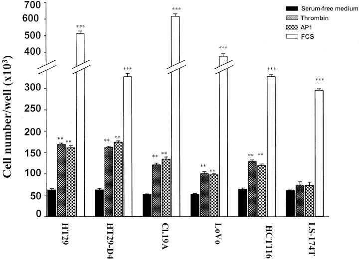
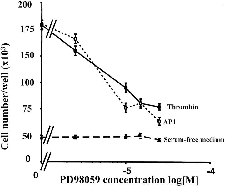
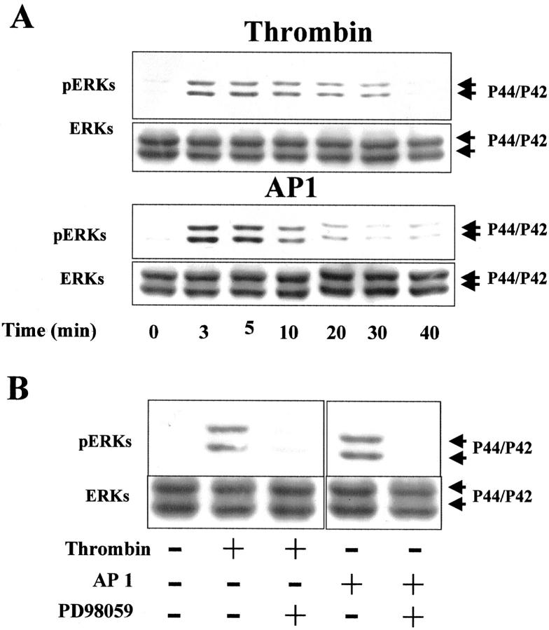
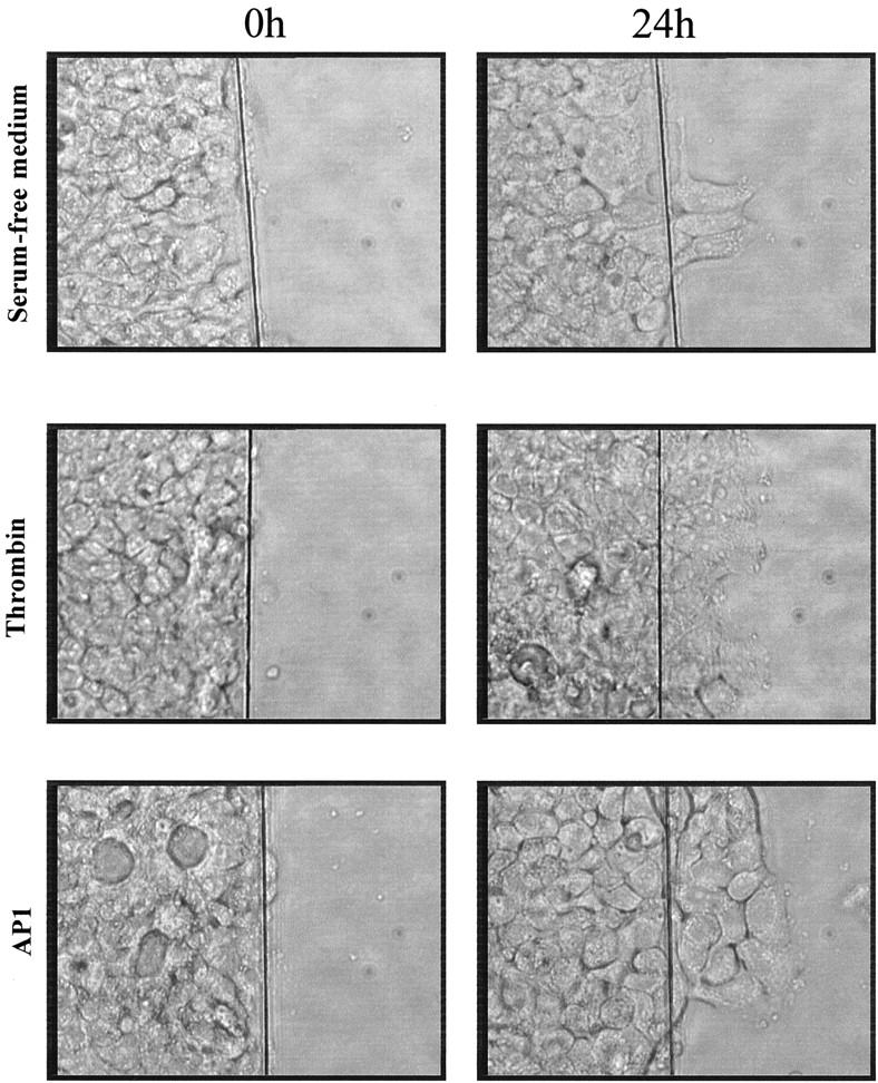
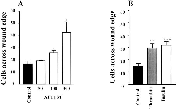
The traditional view on the role of serine proteases in tumor biology has changed with the recent discovery of a family of protease-activated receptors (PARs). In this study we explored the expression and functional role of the thrombin receptor PAR-1 in human colon cancer cells. Reverse transcriptase-polymerase chain reaction analysis showed that PAR-1 mRNAs are present in 11 of 14 human colon cancer cell lines tested but not in normal human colonic epithelial cells. This is in line with the immunolocalization of PAR-1 in human colon tumors and its absence in normal human colonic mucosa. The functional significance of the aberrant expression of PAR-1 in colon cancer cells was then investigated. We found that 1) a prompt increase in intracellular calcium concentration was observed on thrombin (10 nmol/L) or PAR-1 agonist AP1 (100 micro mol/L) challenge of HT29 cells; 2) HT29 quiescent cells treated with thrombin (0.01 to 20 nmol/L) or AP1 (1 to 300 micro mol/L) exhibited dramatic mitogenic responses (3.5-fold increase in cell number). Proliferative effects of thrombin or AP1 were also observed in other colon cancer cell lines expressing PAR-1. This effect was reversed by the MEK inhibitor PD98059 in consonance with the ability of thrombin or AP1 to induce phosphorylation of p42/p44 extracellular-regulated protein kinases. 3) PAR-1 activation by thrombin or AP1 led to a two-fold increase in cell motility of wounded HT29-D4. Our results demonstrate for the first time the aberrant expression of the functional thrombin receptor PAR-1 in colon cancers and its important involvement in cell proliferation and motility. Thrombin should now be considered as a growth factor for human colon cancer.


Figures

Figure 1.
Figure 1.
Constitutive expression of PAR-1 mRNA in various human colon cancer cell lines (A), undifferentiated exponentially growing (day 5) and differentiated postconfluent (day 20) Caco-2 cells (B), and HT29 cells (C) versus normal human colon epithelium. Total RNA extracted was reverse-transcribed and PCR-amplified with PAR-1 (A–C, top), GAPDH primers (A and B, bottom), or PAR-2 primers (C, bottom). A single PCR-amplified product of the exact predicted size (280 bp) for PAR-1 was visualized after electrophoresis in a 2% agarose gel. Note that in B, PAR-1 is present in both undifferentiated and differentiated Caco-2 cells and that in C, contrary to PAR-1, PAR-2 is equally expressed in normal and cancer cells.
Figure 2.
Figure 2.
Immunodetection of PAR-1 receptor in human colon cancer cells with WEDE15 antibody. Left: Immunofluorescence detection of PAR-1. Cells were fixed using 2% paraformaldehyde. PAR-1 protein is evident on the surface of HT29 cells (A), HT29-D4 cells (B), and HCT116 cells (C). No immunofluorescence was observed on LS174T cells (D). Right: Immunoblot of PAR-1 in HT29 cells. One band running at a size of ∼66 kd can be seen. Simultaneous detection with a control IgG does not reveal any immunoreactivity on HT29 lysate. Original magnification, ×400 (A–D).
Figure 3.
Figure 3.
Representative immunostaining for the thrombin receptor PAR-1 with WEDE15 (A–E) in paraffin sections of normal colonic mucosa and colonic tissues from patients with adenocarcinoma. A: PAR-1 immunoreactivity is absent in normal mucosa from control patients. B and C: Two sections from a colonic mucosa distant from a right colon adenocarcinoma. B: Normal mucosa 4 cm from the lesion shows very discrete immunoreactivity on the surface of the epithelium (arrow). C: In the low-grade dysplastic colonic mucosa contiguous to the cancerous lesion, clear immunoreactivity is present in the epithelium of the upper part of the crypt (arrow). D: In the low-grade dysplastic colonic mucosa contiguous to a left colon adenocarcinoma from another patient, similar immunoreactivity as in C is seen. Arrow in the enlarged area in D shows positive immunoreactivity in the colonocytes except in the goblet cells. E: Partial view of the adenocarcinoma contiguous to the dysplastic colonic mucosa seen in D. PAR-1 immunoreactivity in the cancerous epithelial cords is uniformly present but weaker than in the dysplastic mucosa (arrows). Scale bars, 100 μm.
Figure 4.
Figure 4.
Effect of α-thrombin and AP1 on calcium signaling. HT29 cells were loaded with Fura-2/AM and assayed in medium containing Ca2+. A: Thrombin (1 U/ml) followed by a second challenge with trypsin (10 nmol/L). B: The cells were challenged first with the activating peptide AP1 (100 μmol/L) followed by a second challenge with thrombin (1 U/ml) and a third challenge with the activating peptide for PAR-2, AP2 (100 μmol/L). C: HT29 cells were challenged with thrombin (1 U/ml) pretreated with its inhibitor, hirudin (10 U/ml), then with AP1 (100 μmol/L). Note that cells are still responsive to a second challenge with AP1. D: HT29 cells were challenged with AP1 (100 μmol/L) preincubated with hirudin. All of the compounds were given at the arrows. These results are representative of three others.
Figure 5.
Figure 5.
Concentration-dependent stimulation of HT29 cell proliferation by thrombin or AP1. Quiescent cells were grown in serum-free medium with the indicated concentration of thrombin (filled circles), AP1 (open circles), or thrombin and hirudin (10 U/ml) (filled triangle). After 96 hours, cells from triplicate wells were counted for each condition. Data are means ± SD of three different experiments.
Figure 6.
Figure 6.
Ability of PAR-1 ligands (AP1, thrombin) to induce cell proliferation in different human colon cancer cell lines. Quiescent cells were grown for 96 hours in serum-free medium without or with 100 μmol/L of AP1, 10 nmol/L of thrombin, or 10% FCS. Cells from triplicate wells were counted for each condition. Data are means ± SD. ***, P < 0.0001; **, P < 0.001, thrombin-, AP1-, or 10% FCS-treated cells versus control cells.
Figure 7.
Figure 7.
Dose-dependent inhibition of thrombin-induced cell proliferation by the MEK inhibitor PD98059. Quiescent HT29 cells were grown for 96 hours in serum-free medium without (control) or with 100 μmol/L of AP1 or 10 nmol/L of thrombin in the presence of the indicated concentrations of MEK inhibitor (PD98056). Data are means ± SD of three separate experiments.
Figure 8.
Figure 8.
Activation of p42/p44 MAPK by thrombin and AP1 and effect of MEK inhibitor (PD98059) on thrombin- and AP1-induced phosphorylation. A: Immunoblot with phospho-specific p42/p44 MAPK antibodies on HT29 cell lysate treated with or without thrombin (10 nmol/L) or AP1 (100 μmol/L) for the indicated time periods (shown in minutes). B: Phosphorylated form of p42/p44 MAPK in HT29 cells pretreated with 20 μmol/L of PD98059 and then with or without thrombin (10 nmol/L) or AP1 (100 μmol/L) addition for 5 minutes. To confirm equal protein loading, the membranes were stripped and blotted with p42/p44 antibodies.
Figure 9.
Figure 9.
Effect of thrombin on migration of HT29-D4 cells. Wounded monolayers were cultured for 24 hours in serum-free medium in the absence or presence of 5 nmol/L of thrombin or 300 μmol/L of AP1. This result is representative of four independent experiments.
Figure 10.
Figure 10.
Effect of thrombin and AP1 on migration of HT29-D4 cells. The migrating cells across the wound were fixed, stained, and counted. A: Wounded monolayers were cultured for 24 hours in serum-free medium in the presence of various concentration of AP1. B: Wounded monolayers were cultured for 24 hours in serum-free medium in the absence or presence of thrombin (5 nmol/L) or insulin (50 nmol/L) taken as a positive control. Results are means ± SD of three (A) and four (B) independent experiments. ***, P < 0.001; **, P < 0.01; *, P < 0.05, insulin-, thrombin- or AP1-treated cells versus control cells.

References

    1. Chung DC: The genetic basis of colorectal cancer: insights into critical pathways of tumorigenesis. Gastroenterology 2000, 119:854-865 - PubMed
    1. Kinzler KW, Vogelstein B: Lessons from hereditary colorectal cancer. Cell 1996, 87:159-170 - PubMed
    1. Favoni RE, de Cupis A: The role of polypeptide growth factors in human carcinomas: new targets for a novel pharmacological approach. Pharmacol Rev 2000, 52:179-206 - PubMed
    1. Mignatti P, Rifkin DB: Biology and biochemistry of proteinases in tumor invasion. Physiol Rev 1993, 73:161-195 - PubMed
    1. McCawley LJ, Matrisian LM: Matrix metalloproteinases: multifunctional contributors to tumor progression. Mol Med Today 2000, 6:149-156 - PubMed

MeSH terms