

Intermediate cells in human prostate epithelium are enriched in proliferative inflammatory atrophy
- PMID: 12707036
- PMCID: PMC1851184
- DOI: 10.1016/S0002-9440(10)64286-1
Intermediate cells in human prostate epithelium are enriched in proliferative inflammatory atrophy
Abstract
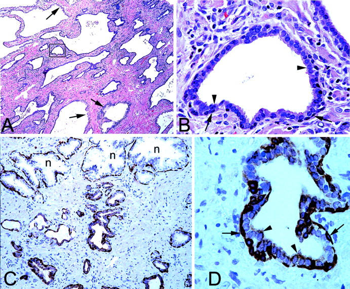
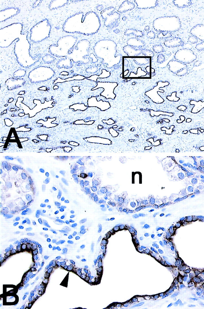
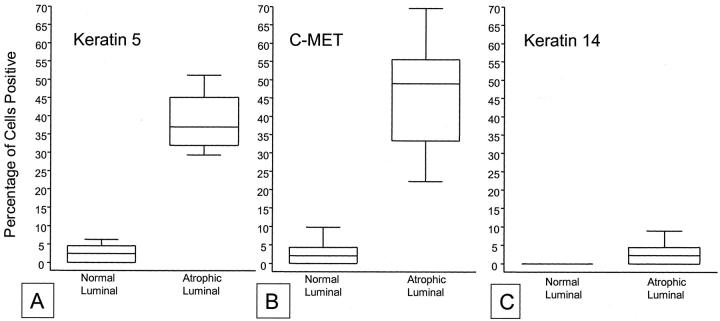
Within the human prostate epithelium four cell populations can be discriminated based on their expression of keratins (K). Basal cells express high levels of K5 and K14, as well as p63, whereas they have very low levels of androgen receptor, prostate-specific antigen (PSA), K8, and K18. Luminal secretory cells lack p63, K5, and K14 but express high levels of K8, K18, androgen receptor, and PSA. Additionally, cells have been identified with a keratin phenotype intermediate between basal and luminal cells that co-express high levels of K5 and K18 (K5/18) as well as hepatocyte growth factor receptor c-MET. Although intermediate cells have been proposed as precursor cells of prostate cancer, their biology is ill defined. Epithelial cells in proliferative inflammatory atrophy (PIA) appear to be cycling rapidly as indicated by expression of Ki-67, and morphological transitions have been identified between PIA and high-grade prostate intraepithelial neoplasia. Many of the atrophic epithelial luminal cells in PIA are candidates for intermediate cells based in part on weak expression of PSA and androgen receptor, high levels of K8/18, and lack of p63. The objective of this study was to further clarify the phenotype of the proposed intermediate cells in PIA and to quantitatively determine the level in which these intermediate cells preferentially occur in PIA lesions. Intermediate cells were immunohistochemically demonstrated using antibodies to K5, K14, K18, and c-MET. Using radical prostatectomy specimens (n = 15) the area fraction of intermediate cells in normally differentiated prostate epithelium and PIA were quantified by a grid point counting method. Atrophic luminal cells of PIA lesions expressed K5 in 39.2 +/- 7.4% of cells compared to 2.4 +/- 2.3% in normal epithelium (P < 0.00001). By contrast, K14 was only expressed in 3.0 +/- 3.2% of the luminal cells. Previous studies have shown that virtually 100% of these atrophic luminal cells are strongly positive for K8/18. c-MET was present in 44.1 +/- 14.1% of luminal cells in PIA but only in 2.1 +/- 2.8% of luminal cells in normal epithelium (P < 0.00001). To unambiguously determine whether intermediate luminal cells in PIA show increased proliferative activity and decreased p27(kip1) expression, double-staining immunofluorescence of Ki-67 and K5, as well as p27(Kip1) and K5 was performed. Luminal cells in PIA often co-expressed K5 and Ki-67. Although p27(Kip1) was strongly expressed in K5-negative differentiated cells in normal epithelium, p27(Kip1) staining was absent in many of the K5-positive cells in the luminal compartment of PIA. We conclude that cells phenotypically intermediate between basal and secretory cells are enriched in PIA lesions. The finding of a large number of highly proliferating intermediate cells in PIA provides further support that these cells may serve as preferred target cells in prostate carcinogenesis.
Figures

References
-
- Potten CS, Morris RJ: Epithelial stem cells in vivo. J Cell Sci 1988, 10:S45-S62 - PubMed
-
- Potten CS, Loeffler M: Stem cells: attributes, cycles, spirals, pitfalls and uncertainties. Lessons for and from the crypt. Development 1990, 110:1001-1020 - PubMed
-
- De Marzo AM, Nelson WG, Meeker AK, Coffey DS: Stem cell features of benign and malignant prostate epithelial cells. J Urol 1998, 160:2381-2392 - PubMed
-
- van Leenders GJ, Schalken JA: Stem cell differentiation within the human prostate epithelium: implications for prostate carcinogenesis. BJU Int 2001, 88(Suppl 2):35-50 - PubMed
-
- Evans GS, Chandler JA: Cell proliferation studies in rat prostate. I. The proliferative role of basal and secretory epithelial cells during normal growth. Prostate 1987, 10:163-178 - PubMed
Publication types
MeSH terms
Substances
Grants and funding
LinkOut - more resources
Full Text Sources
Other Literature Sources
Medical
Research Materials
Miscellaneous
