. 2003 May;162(5):1661-7.
doi: 10.1016/S0002-9440(10)64300-3.

Identification and characterization of the hepatic stellate cell transferrin receptor

Affiliations

Identification and characterization of the hepatic stellate cell transferrin receptor

Kim R Bridle et al. Am J Pathol. 2003 May.

Abstract

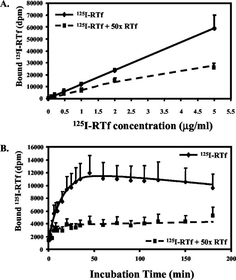
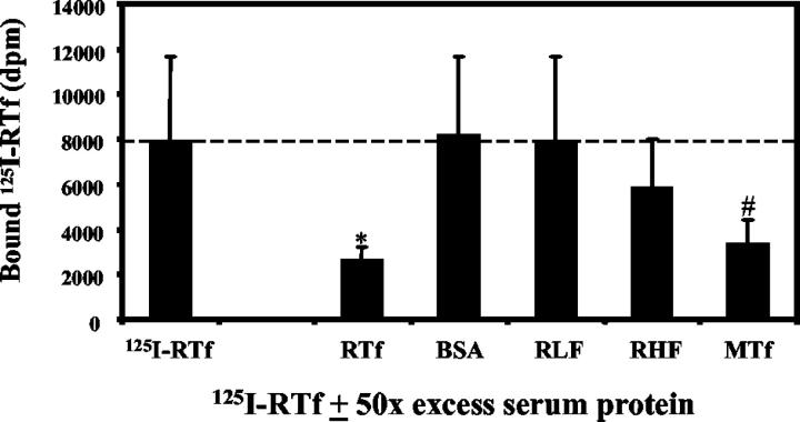
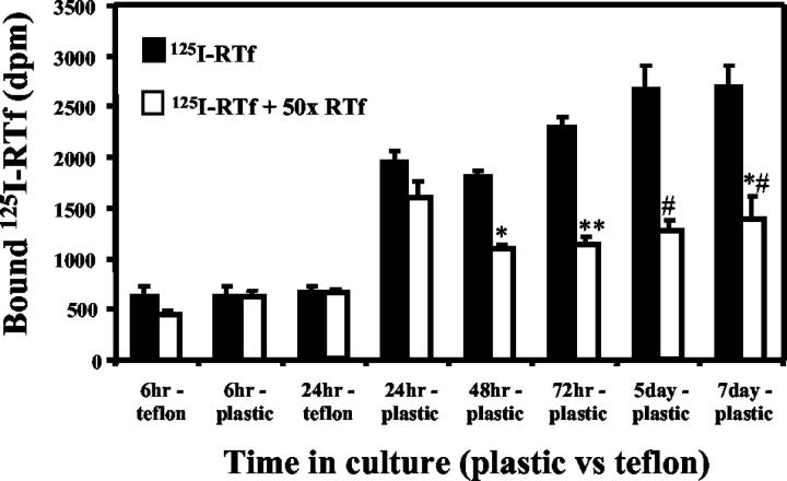
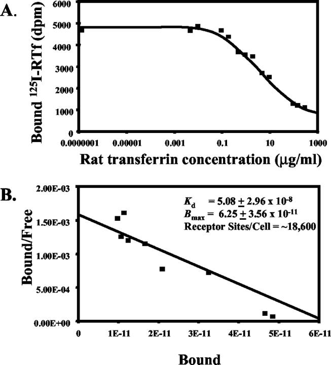
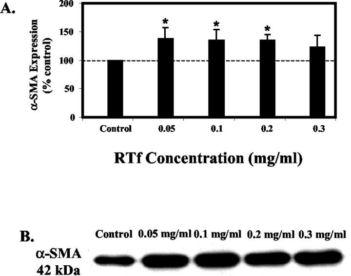
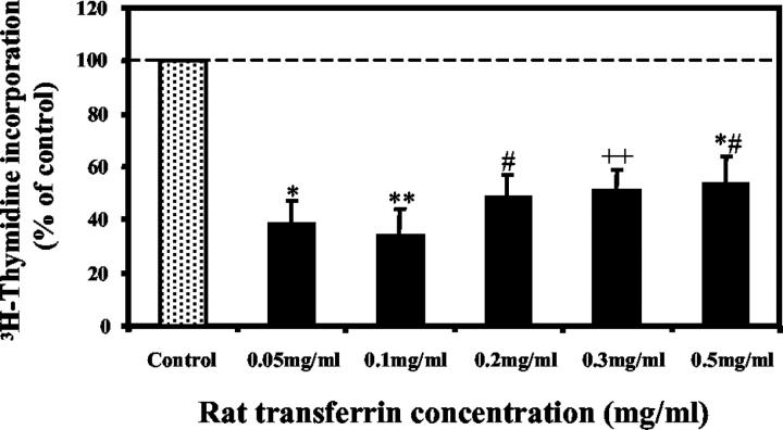
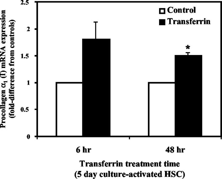
Activated hepatic stellate cells have been implicated in the fibrogenic process associated with iron overload, both in animal models and in human hemochromatosis. Previous studies have evaluated the role of ferritin/ferritin receptor interactions in the activation of stellate cells and subsequent fibrogenesis; however, the role of transferrin in hepatic stellate cell biology is unknown. This study was designed to identify and characterize the stellate cell transferrin receptor and to evaluate the influence of transferrin on stellate cell activation. Identification and characterization of the stellate cell transferrin receptor was determined by competitive displacement assays. The effect of transferrin on stellate cell activation was assessed using western blot analysis for alpha-smooth muscle actin expression, [(3)H]Thymidine incorporation, and real-time RT-PCR for procollagen alpha1(I) mRNA expression. A specific receptor for rat transferrin was observed on activated but not quiescent stellate cells. Transferrin significantly increased the expression of alpha-smooth muscle actin, but caused a decrease in proliferation. Transferrin induced a significant increase in procollagen alpha1(I) mRNA expression. In conclusion, this study has demonstrated for the first time a specific, high affinity receptor for rat transferrin on activated hepatic stellate cells, which via interaction with transferrin regulates stellate cell activation. This suggests that transferrin may be an important factor in the activation of hepatic stellate cells in conditions of iron overload.


Figures

Figure 1.
Figure 1.
A: Optimization of 125I-RTf concentration. HSC were incubated with increasing concentrations of 125I-RTf ± 50-fold excess of unlabelled RTf at 37°C for 2 hours. Optimal specific binding was achieved using 0.5 μg/ml 125I-RTf (59.9 ± 7.3%). Mean ± SE, n = 2, DPM = disintegrations per minute. B: Time course of 125I-RTf uptake by HSC. HSC were incubated with 0.5 μg/ml 125I-RTf ± 50-fold excess of unlabeled RTf at 37°C for up to 180 minutes. Binding reached saturation at ∼45 minutes. Mean ± SE, n = 3. DPM, disintegrations per minute.
Figure 2.
Figure 2.
Identification of the HSC transferrin receptor. HSC were incubated with 0.5 μg/ml 125I-RTf ± 50-fold excess of either unlabelled RTf, BSA, RLF, RHF or MTf at 37°C for 2 hours. HSC demonstrated 67.4 ± 19.3% specific binding for RTf. Mean ± SE, n = 3. *P = 0.01, #P = 0.02 when compared to 125I-RTf. DPM, disintegrations per minute. MTf, mouse transferrin; BSA, bovine serum albumin; RLF, rat liver ferritin; RHF, rat heart ferritin.
Figure 3.
Figure 3.
Competitive binding assay in quiescent and activated HSC. HSC were incubated at 37°C for 2 hours with 0.5 μg/ml 125I-RTf ± 50-fold excess of unlabelled RTf. Cells were cultured on Teflon inserts (to maintain HSC in a quiescent phenotype) and on tissue culture plastic (to activate HSC). 125I-RTf uptake was examined at 6, 24, 48, and 72 hours, and 5 and 7 days post-isolation. Quiescent HSC did not express Tf receptors; however, those cultured on plastic, and therefore culture-activated, did demonstrate competitive displacement of 125I-RTf by a 50-fold excess of unlabelled Tf. Mean ± SE, n = 3. *P = 0.007, **P = 0.003, #P = 0.01, ##P = 0.05, when compared to 125I-RTf. DPM, disintegrations per minute.
Figure 4.
Figure 4.
Scatchard analysis of the competitive displacement of 125I-RTf by unlabeled RTf by the HSC transferrin receptor. A: HSC were incubated with 0.5 μg/ml 125I-RTf ± increasing concentrations of unlabelled RTf at 37°C for 2 hours. B: Scatchard analysis indicated a single class of binding sites for RTf with an estimated Kd of 5.08 ± 2.96 × 10⁻⁸ and a Bmax of 6.25 ± 3.56 × 10⁻¹¹, with approximately 18,600 receptor sites per cell. Results are expressed as means from duplicate determinations in each experiment from HSC isolated from two separate rats.
Figure 5.
Figure 5.
Expression of α-SMA in HSC. A: HSC were treated with increasing concentrations of RTf and collected after 7 days in culture and quantitated using scanning laser densitometry following Western blotting. (B) Representative Western blot of HSC cell extracts (10 μg cell protein/lane) showing 42 kd band corresponding to α-SMA. Results are mean ± SE, n = 4. *P < 0.05 when compared to control.
Figure 6.
Figure 6.
Effect of RTf on HSC proliferation as determined by [3H]thymidine incorporation. Results are expressed as a percentage of control and calculated as disintegrations/min. Results are mean ± SE of quadruplicate determinations in each experiment from HSC isolated from two separate rats. *P = 0.002, **P = 0.001, #P = 0.009, ##P = 0.01, *#P = 0.03, when compared to controls.
Figure 7.
Figure 7.
Effect of RTf on HSC procollagen α1 (I) mRNA expression as determined by real-time RT-PCR analysis. Day 5 HSC were treated with 0.2 mg/ml RTf. RNA was collected at 6 and 48 hours after treatment with RTf. Procollagen α1(I) mRNA expression in RTF-treated HSC are expressed as a fold-difference from controls (untreated HSC), after standardization against expression of the housekeeping gene β-actin. *P = 0.005. Mean ± SE, n = 3.

References

    1. Friedman SL: Hepatic stellate cells. Prog Liver Dis 1996, 14:101-130 - PubMed
    1. Ooi LP, Crawford DH, Gotley DC, Clouston AD, Strong RW, Gobe GC, Halliday JW, Bridle KR, Ramm GA: Evidence that “myofibroblast-like” cells are the cellular source of capsular collagen in hepatocellular carcinoma. J Hepatol 1997, 26:798-807 - PubMed
    1. Ramm GA, Nair VG, Bridle KR, Shepherd RW, Crawford DH: Contribution of hepatic parenchymal and nonparenchymal cells to hepatic fibrogenesis in biliary atresia. Am J Pathol 1998, 153:527-535 - PMC - PubMed
    1. Ramm GA, Crawford DH, Powell LW, Walker NI, Fletcher LM, Halliday JW: Hepatic stellate cell activation in genetic haemochromatosis. Lobular distribution, effect of increasing hepatic iron and response to phlebotomy. J Hepatol 1997, 26:584-592 - PubMed
    1. Ramm GA, Li SC, Li L, Britton RS, O’Neill R, Kobayashi Y, Bacon BR: Chronic iron overload causes activation of rat lipocytes in vivo. Am J Physiol 1995, 268:G451-G458 - PubMed

Publication types

LinkOut - more resources