Comment
. 2003 Apr 21;197(8):955-66.
doi: 10.1084/jem.20021024.

Topological requirements and signaling properties of T cell-activating, anti-CD28 antibody superagonists

Affiliations
Comment

Topological requirements and signaling properties of T cell-activating, anti-CD28 antibody superagonists

Fred Lühder et al. J Exp Med. .

Abstract

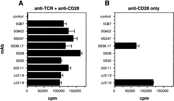
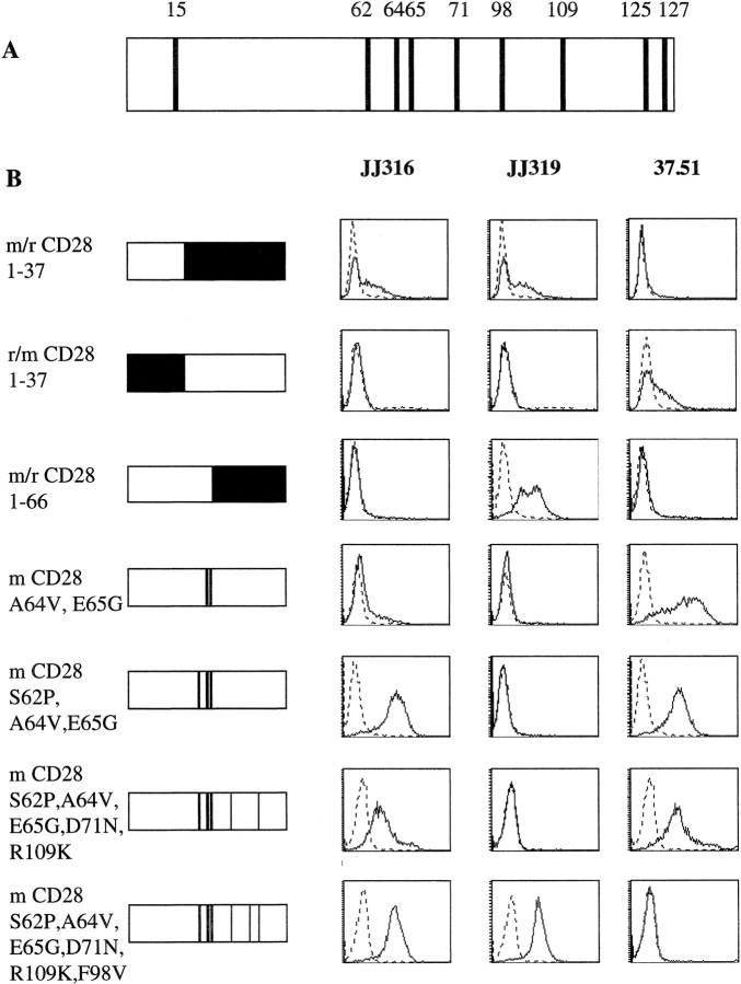
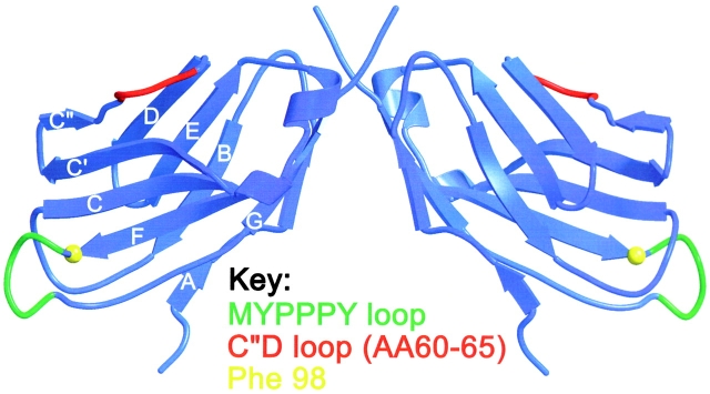
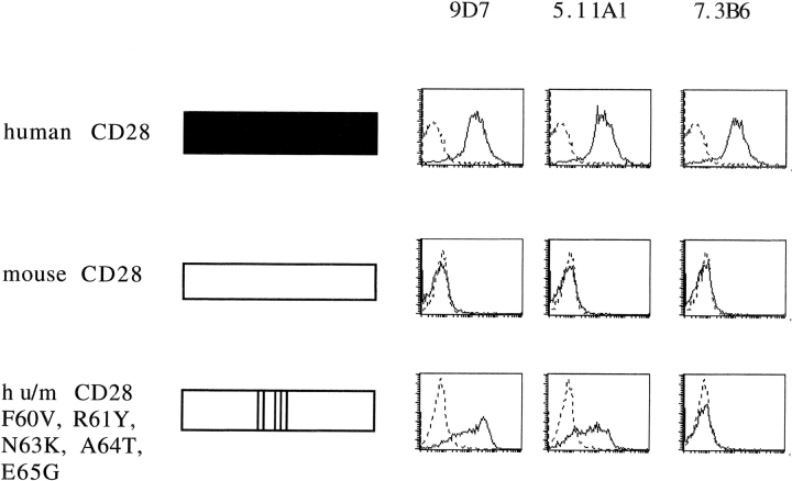
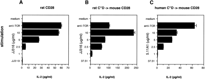
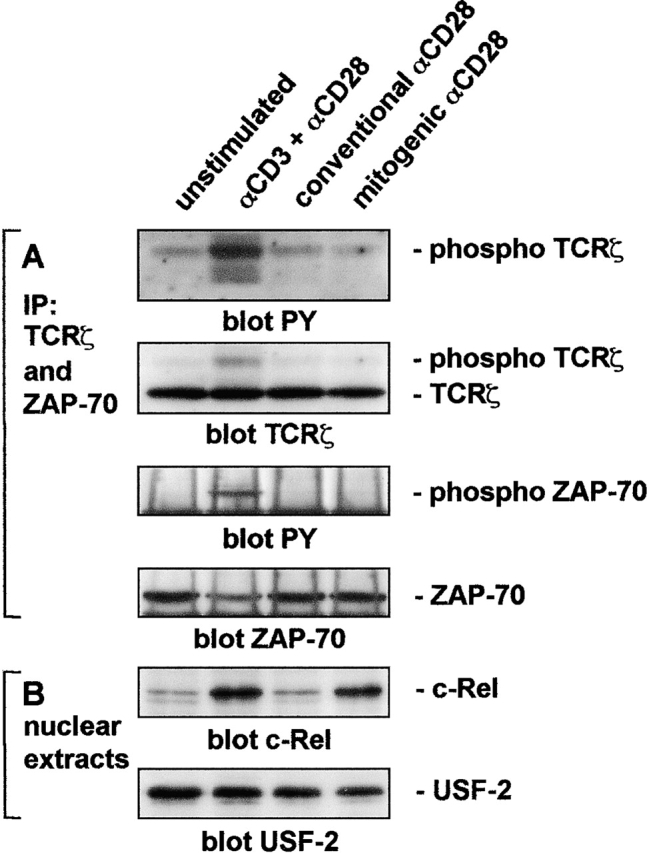
Full activation of naive T cells requires both engagement of the T cell antigen receptor (TCR; signal 1) and costimulatory signaling by CD28 (signal 2). We previously identified two types of rat CD28-specific monoclonal antibodies (mAbs): "conventional," TCR signaling-dependent costimulatory mAbs and "superagonistic" mAbs capable of inducing the full activation of primary resting T cells in the absence of TCR ligation both in vitro and in vivo. Using chimeric rat/mouse CD28 molecules, we show that the superagonists bind exclusively to the laterally exposed C"D loop of the immunoglobulin-like domain of CD28 whereas conventional, costimulatory mAbs recognize an epitope close to the binding site for the natural CD80/CD86 ligands. Unexpectedly, the C"D loop reactivity of a panel of new antibodies raised against human CD28 could be predicted solely on the basis of their superagonistic properties. Moreover, mouse CD28 molecules engineered to express the rat or human C"D loop sequences activated T cell hybridomas without TCR ligation when cross-linked by superagonistic mAbs. Finally, biochemical analysis revealed that superagonistic CD28 signaling activates the nuclear factor kappaB pathway without inducing phosphorylation of either TCRzeta or ZAP70. Our findings indicate that the topologically constrained interactions of anti-CD28 superagonists bypass the requirement for signal 1 in T cell activation. Antibodies with this property may prove useful for the development of T cell stimulatory drugs.


Figures

Figure 1.
Figure 1.
Costimulation and direct stimulation of primary rat T cells by rat CD28-specific mAb. (A) Nylon wool–purified rat lymph node T cells (105/well) were stimulated by anti-TCR mAb R73 immobilized via sheep anti–mouse IgG and soluble rat CD28-specific mAb. An isotype-matched irrelevant mAb served as negative control for the CD28-specific mAb. On day 3, the cells were pulsed with 0.5 μCi [3H]thymidine per well for 16 h followed by harvesting and β counting. (B) As in A but without anti-TCR mAb. One representative experiment from two with the identical outcome is shown.
Figure 2.
Figure 2.
Epitope mapping of a superagonistic (JJ316) and conventional (JJ319) rat CD28-specific mAb. (A) Comparison of extracellular portions of mouse and rat CD28. Positions of aa differences are indicated by numbers. (B) Binding of superagonistic and conventional mAbs to rat/mouse CD28 chimeras. Mouse sequences are represented in white and rat sequences in black. Positions of swapped aa are indicated on the left. All constructs were stably expressed in L929 fibroblast cells. FACS® histograms are shown for superagonistic (JJ316) and conventional (JJ319) rat and for the conventional mouse CD28-specific mAb 37.51 (solid lines) and an isotype-matched control mAb (dotted lines). At least two independent experiments were performed and a representative experiment is shown.
Figure 3.
Figure 3.
Depiction of the epitopes for superagonistic and conventional CD28-specific mAbs in a three-dimensional model of the extracellular part of human CD28. The model of the CD28 monomer was derived by computer calculation from the X ray crystallographic structure of murine CTLA-4 using the sequence alignment derived by Metzler et al. (reference 25). The dimer was constructed on the basis of the CTLA-4 homodimer observed in the crystals of the complex of CTLA-4 and CD80, and is shown with the membrane-proximal region at the top. The MYPPPY motif (aa 99–104) critical for B7 binding is indicated in green, the adjacent aa 98 residue critical for binding of the conventional rat and mouse CD28-specific mAb is highlighted in yellow, and the C′′D loop responsible for the binding of superagonistic rat and human CD28-specific mAb (aa 60–65) is indicated in red.
Figure 4.
Figure 4.
Stimulation of human T cells by superagonistic anti-CD28 mAb in the presence and absence of TCR triggering. Freshly isolated human T cells from PBMC were cultured at 5 × 10⁵/ml in plates containing (A) or lacking (B) immobilized anti-CD3 mAb (suboptimal concentration of 0.003 μg/ml) and soluble anti-CD28 mAb7.3B6 (a representative costimulatory mAb), 9D7 or 5.11A1 (3 μg/ml; two novel superagonistic mAb). (C) Titration of anti-CD28 mAb in the absence of anti-CD3 mAb. (A–C) Cells were cultured for 3 d before the addition of [3H]thymidine. Data are representative for four (A and B) or two (C) independent experiments.
Figure 5.
Figure 5.
Epitope mapping of superagonistic human CD28-specific mAb. Binding of two superagonistic (9D7 and 5.11A1) and a conventional (7.3B6) human CD28-specific mAb to L 929 fibroblast cells expressing full-length human CD28 (top), full-length mouse CD28 (middle), and a chimeric human/mouse CD28 molecule with a humanized C”D loop (bottom). Human sequences are shown in black and mouse sequences in white. FACS® histograms are shown for 9D7, 5.11A1, and 7.3B6 (solid lines), and for an isotype-matched control mAb (dotted lines). At least two independent experiments were performed and a representative experiment is shown.
Figure 6.
Figure 6.
Activation of T cell hybridoma cells via superagonistic CD28-specific mAb. The 58 T cell hybridoma line supplemented with a rat TCR was retrovirally transduced with mouse CD28 containing the following replacements: (A) the whole extracellular part of rat CD28, (B) the rat C′′D loop, or (C) the human C′′D loop. The cell lines were left unstimulated or were stimulated via immobilized anti-TCR mAb R73, superagonistic CD28-specific mAb (JJ316 for rat, 5.11A1 for human) at the indicated final concentrations, ranging from 10 to 1 μg/ml, or conventional CD28-specific mAb (JJ319 for rat, 37.51 for the chimeric molecules). After 2 d, supernatants were tested for the presence of mouse IL-2 by ELISA. Because stimulation with conventional CD28-specific mAb did not result in substantial IL-2 production at any concentration tested, only results using the highest concentration (10 μg/ml) are presented. One of at least three independent experiments with similar results is shown.
Figure 7.
Figure 7.
Mitogenic anti-CD28 antibody stimulation activates NF-κB without inducing tyrosine phosphorylation of TCRζ or ZAP-70. (A) T cells were preincubated with the indicated stimulating antibodies for 1 h at 4°C, washed, incubated with 10 μg/ml rat anti–mouse IgG for 30 min at 4°C, and incubated for 0.5 min at 37°C before the addition of NP-40 lysis buffer. Proteins were precipitated with antibodies to ZAP-70 and TCRζ precoupled to protein G–Sepharose, resolved by SDS-PAGE, Western blotted, and probed with antiphosphotyrosine antibody. Membranes were reprobed with antibodies to TCRζ and ZAP-70 to ensure comparable loading. (B) Nuclear extracts from the same cells stimulated with the same primary antibodies and incubated for 20 h on sheep anti–mouse IgG-coated plates were resolved by SDS-PAGE, Western blotted, and probed with antibodies to c-Rel or USF-2 as a loading control.

Comment on

  • CD28, costimulator or agonist receptor?
    Margulies DH. Margulies DH. J Exp Med. 2003 Apr 21;197(8):949-53. doi: 10.1084/jem.20030303. J Exp Med. 2003. PMID: 12707298 Free PMC article. No abstract available.

References

    1. Schwartz, R.H. 1990. A cell culture model for T lymphocyte clonal anergy. Science. 248:1349–1356. - PubMed
    1. Sharpe, A.H., and G.J. Freeman. 2002. The B7-CD28 superfamily. Nat. Rev. Immunol. 2:116–126. - PubMed
    1. Rudd, C.E. 1996. Upstream-downstream: CD28 cosignaling pathways and T cell function. Immunity. 4:527–534. - PubMed
    1. Viola, A., S. Schroeder, Y. Sakakibara, and A. Lanzavecchia. 1999. T lymphocyte costimulation mediated by reorganization of membrane microdomains. Science. 283:680–682. - PubMed
    1. Wulfing, C., and M.M. Davis. 1998. A receptor/cytoskeletal movement triggered by costimulation during T cell activation. Science. 282:2266–2269. - PubMed

Publication types

MeSH terms