. 2003 Apr 21;197(8):1059-63.
doi: 10.1084/jem.20030240.

Human dendritic cells activated by TSLP and CD40L induce proallergic cytotoxic T cells

Affiliations

Human dendritic cells activated by TSLP and CD40L induce proallergic cytotoxic T cells

Michel Gilliet et al. J Exp Med. .

Abstract

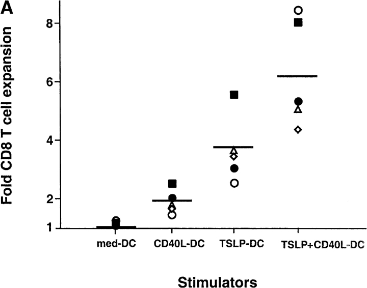
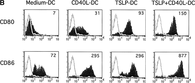
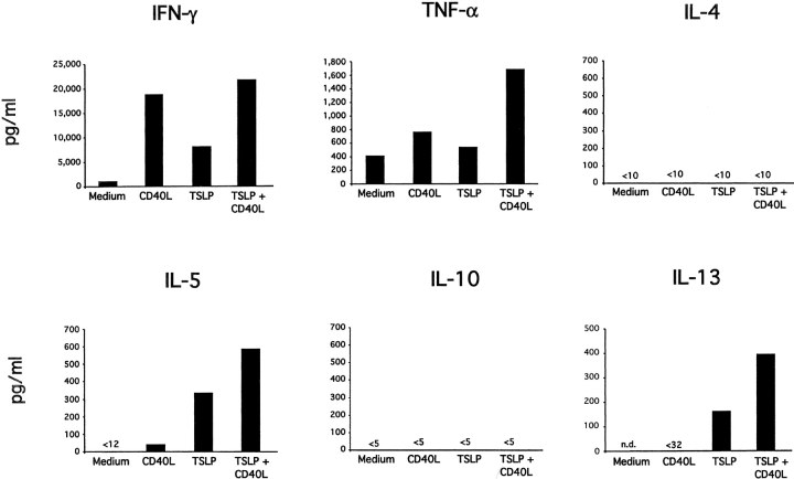
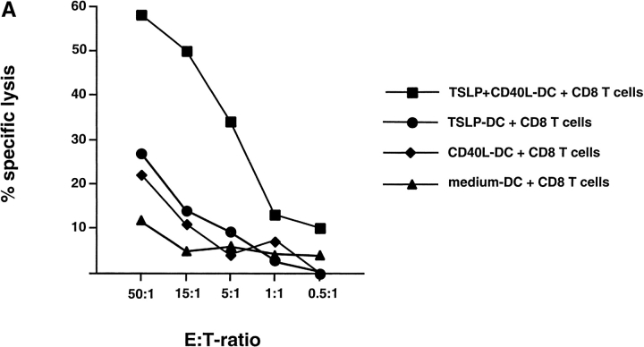
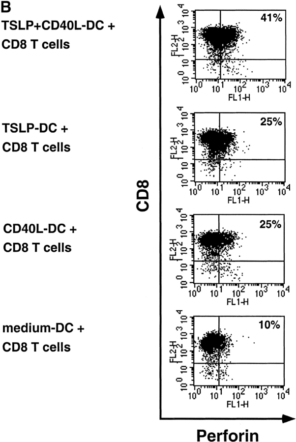
Human thymic stromal lymphopoietin (TSLP) is a novel epithelial cell-derived cytokine, which induces dendritic cell (DC)-mediated CD4+ T cell responses with a proallergic phenotype. Although the participation of CD8+ T cells in allergic inflammation is well documented, their functional properties as well as the pathways leading to their generation remain poorly understood. Here, we show that TSLP-activated CD11c+ DCs potently activate and expand naive CD8+ T cells, and induce their differentiation into interleukin (IL)-5 and IL-13-producing effectors exhibiting poor cytolytic activity. Additional CD40L triggering of TSLP-activated DCs induced CD8+ T cells with potent cytolytic activity, producing large amounts of interferon (IFN)-gamma, while retaining their capacity to produce IL-5 and IL-13. These data further support the role of TSLP as initial trigger of allergic T cell responses and suggest that CD40L-expressing cells may act in combination with TSLP to amplify and sustain pro-allergic responses and cause tissue damage by promoting the generation of IFN-gamma-producing cytotoxic effectors.


Figures

Figure 1.
Figure 1.
CD11c+ DCs activated by TSLP alone or in combination with CD40L induce a strongly expansion of naive CD8+ T cells. (A) Naive CD8+ T cells were cultured for 6 d with CD11c+ DCs activated for 24 h with medium, CD40L, TSLP, or TSLP + CD40. TSLP-DCs induced stronger expansion of naive CD8+ T cells than CD40-L-DCs. The combination of TSLP and CD40L showed an additive effect on the capacity of activated DCs to induce CD8+ T cell expansion. Data represent five independent experiments; horizontal bars represent the median value. (B) Surface expression of CD80 and CD86 by medium-DCs, CD40L-DCs, TSLP-DCs, and TSLP+CD40L-DCs. TSLP-DCs showed significantly higher levels of CD80 than CD40L-DCs, TSLP+CD40L-DCs showed the highest levels of both CD80 and CD86. Filled histograms represent staining of costimulatory molecules CD80 and CD86; open histograms represent the isotype control. Numbers indicate the mean fluorescence intensity.
Figure 1.
Figure 1.
CD11c+ DCs activated by TSLP alone or in combination with CD40L induce a strongly expansion of naive CD8+ T cells. (A) Naive CD8+ T cells were cultured for 6 d with CD11c+ DCs activated for 24 h with medium, CD40L, TSLP, or TSLP + CD40. TSLP-DCs induced stronger expansion of naive CD8+ T cells than CD40-L-DCs. The combination of TSLP and CD40L showed an additive effect on the capacity of activated DCs to induce CD8+ T cell expansion. Data represent five independent experiments; horizontal bars represent the median value. (B) Surface expression of CD80 and CD86 by medium-DCs, CD40L-DCs, TSLP-DCs, and TSLP+CD40L-DCs. TSLP-DCs showed significantly higher levels of CD80 than CD40L-DCs, TSLP+CD40L-DCs showed the highest levels of both CD80 and CD86. Filled histograms represent staining of costimulatory molecules CD80 and CD86; open histograms represent the isotype control. Numbers indicate the mean fluorescence intensity.
Figure 2.
Figure 2.
TSLP-activated DCs induce IL-5 and IL-13–producing CD8+ T cells. Cytokine production by naive CD8+ T cells primed for 6 d with medium-DCs, CD40L-DCs, TSLP-DCs, or TSLP+CD40L-DCs. Harvested CD8+ T cells were restimulated for 24 h with anti-CD3 and anti-CD28 and IFN-γ, TNF-α, IL-4, IL-5, IL-10, and IL-13 were measured in the culture supernatant by ELISA. TSLP-DCs and TSLP+CD40L-DCs both induced IL-5 and IL-13 producing CD8+ T cells. However, in contrast to TSLP-DCs, TSLP+CD40L-DCs primed CD8+ T effector cells to produce high levels of IFN-γ. Data represent one of three independent experiments.
Figure 3.
Figure 3.
CD40L-TSLP-DCs induced the generation of CD8+ cytotoxic effector T cells. (A) Naive CD8+ T cells were primed for 6 d with medium-DCs (triangles), CD40L-DCs (diamonds), TSLP-DCs (circles), or TSLP+CD40L-DCs (squares), collected and tested for their ability to lyse allogeneic HLA-A2–positive target cells. Specific lysis of autologous HLA-A2–negative target cells remained below 15%. Whereas both TSLP-DCs and CD40L-DCs induced CD8+ T cells with poor cytolytic activity, TSLP+CD40L-DCs induced CD8+ T cells with potent cytolytic activity. Data represent one of three independent experiments. (B) Two color staining for intracellular perforin and surface CD8 of T cells primed with medium-DCs, CD40-L-DCs, TSLP-DCs, or TSLP+CD40-L-DCs for 6 d.
Figure 3.
Figure 3.
CD40L-TSLP-DCs induced the generation of CD8+ cytotoxic effector T cells. (A) Naive CD8+ T cells were primed for 6 d with medium-DCs (triangles), CD40L-DCs (diamonds), TSLP-DCs (circles), or TSLP+CD40L-DCs (squares), collected and tested for their ability to lyse allogeneic HLA-A2–positive target cells. Specific lysis of autologous HLA-A2–negative target cells remained below 15%. Whereas both TSLP-DCs and CD40L-DCs induced CD8+ T cells with poor cytolytic activity, TSLP+CD40L-DCs induced CD8+ T cells with potent cytolytic activity. Data represent one of three independent experiments. (B) Two color staining for intracellular perforin and surface CD8 of T cells primed with medium-DCs, CD40-L-DCs, TSLP-DCs, or TSLP+CD40-L-DCs for 6 d.

References

    1. Sims, J.E., D.E. Williams, P.J. Morrissey, K. Garka, D. Foxworthe, V. Price, S.L. Friend, A. Farr, M.A. Bedell, N.A. Jenkins, et al. 2000. Molecular cloning and biological characterization of a novel murine lymphoid growth factor. J. Exp. Med. 192:671–680. - PMC - PubMed
    1. Reche, P.A., V. Soumelis, D.M. Gorman, T. Clifford, M. Liu, M. Travis, S.M. Zurawski, J. Johnston, Y.J. Liu, H. Spits, et al. 2001. Human thymic stromal lymphopoietin preferentially stimulates myeloid cells. J. Immunol. 167:336–343. - PubMed
    1. Soumelis, V., P.A. Reche, H. Kanzler, W. Yuan, G. Edward, B. Homey, M. Gilliet, S. Ho, S. Antonenko, A. Lauerma, et al. 2002. Human epithelial cells trigger dendritic cell mediated allergic inflammation by producing TSLP. Nat. Immunol. 3:673–680. - PubMed
    1. Kay, A.B. 2001. Allergy and allergic diseases. First of two parts. N. Engl. J. Med. 344:30–37. - PubMed
    1. Renauld, J.C. 2001. New insights into the role of cytokines in asthma. J. Clin. Pathol. 54:577–589. - PMC - PubMed

Publication types

MeSH terms