. 2003 Apr 28;161(2):403-16.
doi: 10.1083/jcb.200303036. Epub 2003 Apr 21.

Defining desmosomal plakophilin-3 interactions

Affiliations

Defining desmosomal plakophilin-3 interactions

Stefan Bonné et al. J Cell Biol. .

Abstract

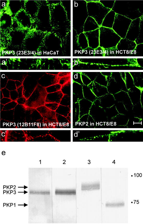
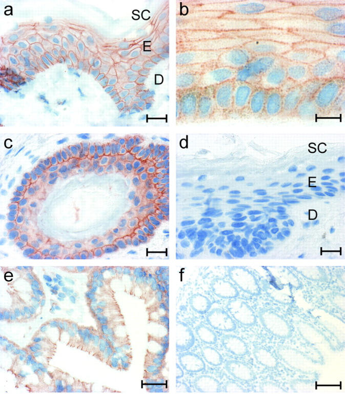
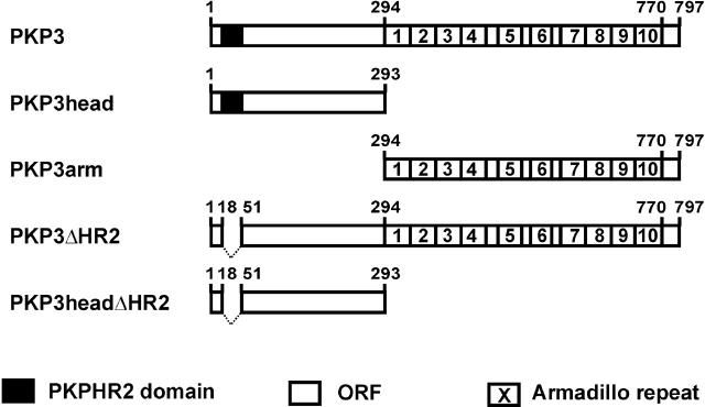
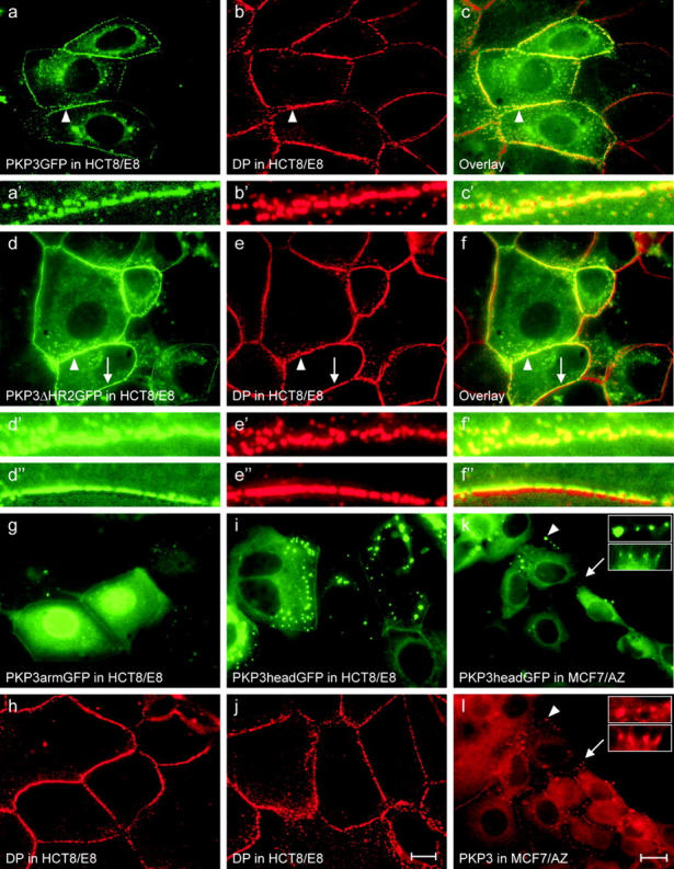
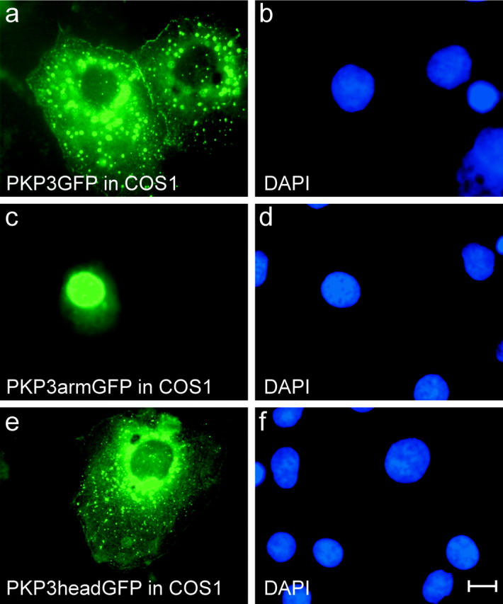
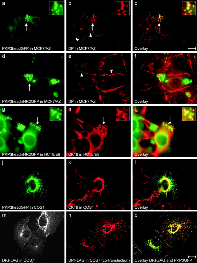
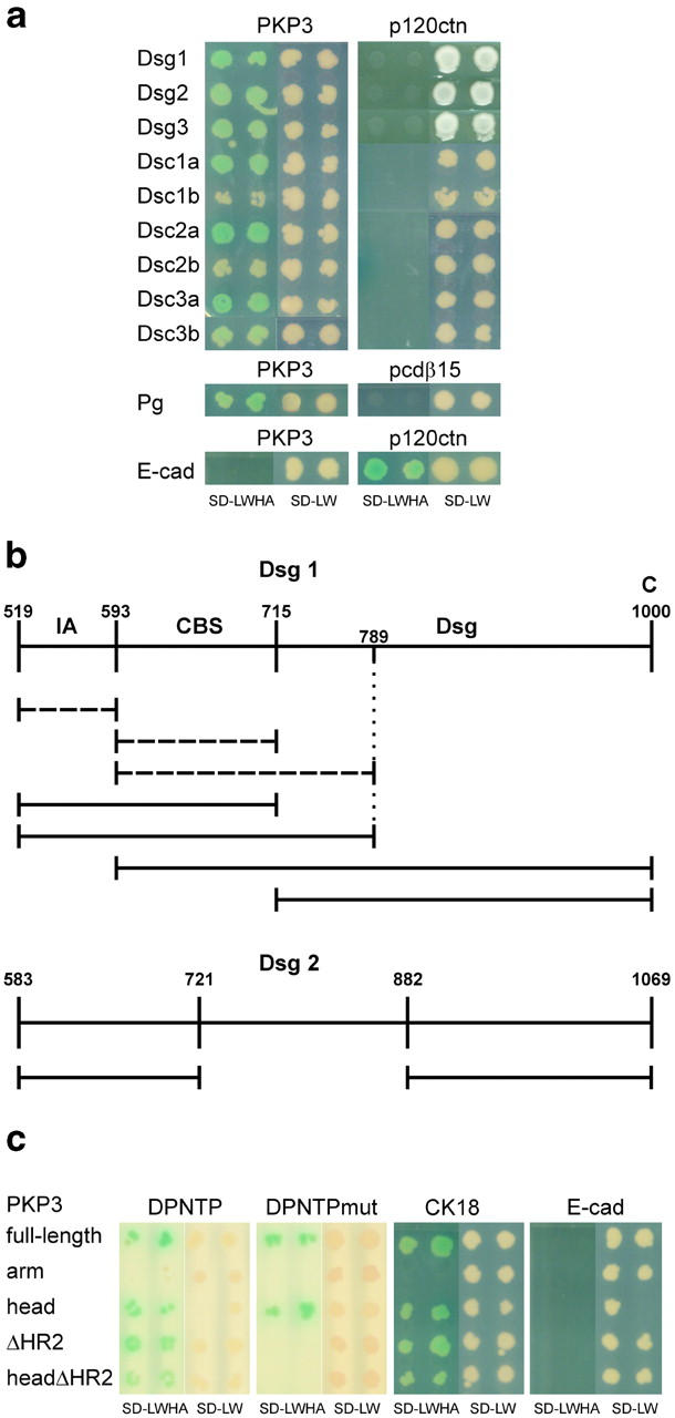
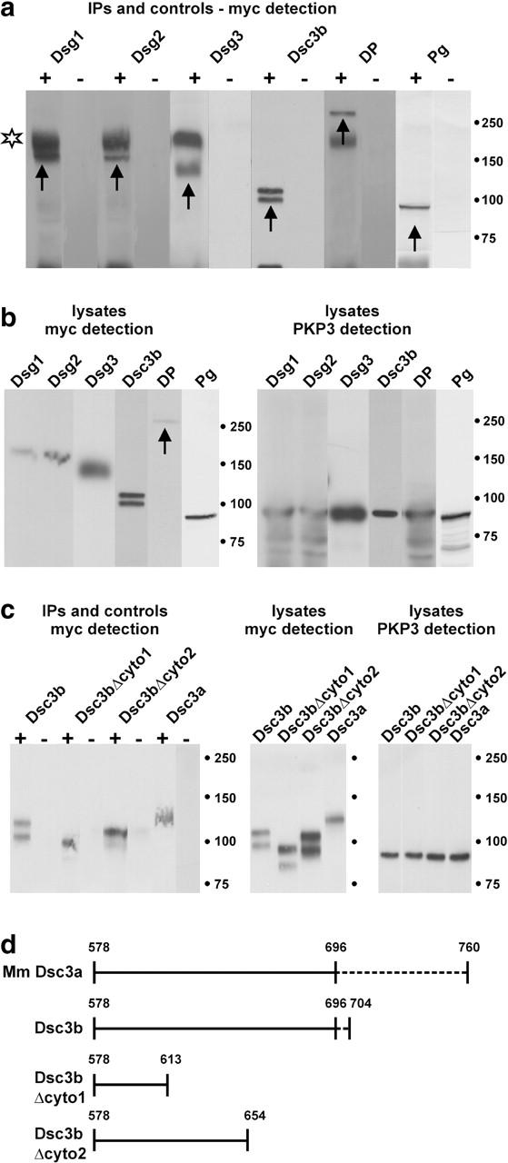
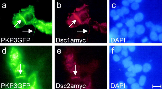
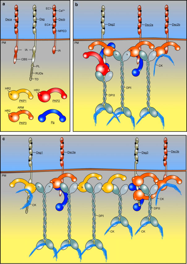
Plakophilin 3 (PKP3) is a recently described armadillo protein of the desmosomal plaque, which is synthesized in simple and stratified epithelia. We investigated the localization pattern of endogenous and exogenous PKP3 and fragments thereof. The desmosomal binding properties of PKP3 were determined using yeast two-hybrid, coimmunoprecipitation and colocalization experiments. To this end, novel mouse anti-PKP3 mAbs were generated. We found that PKP3 binds all three desmogleins, desmocollin (Dsc) 3a and -3b, and possibly also Dsc1a and -2a. As such, this is the first protein interaction ever observed with a Dsc-b isoform. Moreover, we determined that PKP3 interacts with plakoglobin, desmoplakin (DP) and the epithelial keratin 18. Evidence was found for the presence of at least two DP-PKP3 interaction sites. This finding might explain how lateral DP-PKP interactions are established in the upper layers of stratified epithelia, increasing the size of the desmosome and the number of anchoring points available for keratins. Together, these results show that PKP3, whose epithelial and epidermal desmosomal expression pattern and protein interaction repertoire are broader than those of PKP1 and -2, is a unique multiprotein binding element in the basic architecture of a vast majority of epithelial desmosomes.


Figures

Figure 1.
Figure 1.
Immunodetection of PKPs using different mouse mAbs. PKP3, detected with mAb 23E3/4 (a and b), 12B11F8 (c), or PKP2 (d) show a similar expression pattern along cell–cell contacts in methanol-fixed HaCaT (a) and HCT8/E8 (b–d) cells. Larger magnifications display the punctate localization of these proteins along cell–cell contacts (a′–d′). Bar, 10 μm. (e) Western blot detection of PKP3 (lane 1, mAb 12B11F8; lane 2, 23E3/4), PKP2 (lane 3), and PKP1 (lane 4) in HaCaT protein lysates. Equal amounts of protein were loaded in each lane.
Figure 2.
Figure 2.
Immunohistochemical detection of PKP3 in paraffin sections of formalin-fixed human skin and colon using mAb 23E3/4. PKP3 is expressed in the living cell layers of the epidermis, but not in the stratum corneum and the dermis (a). At higher magnification, the interrupted PKP3 localization along cell–cell contacts is visible (b). The epidermal cells of the hair root also express PKP3 (c); no signal can be detected in the negative control sections, in which the primary antibody was omitted (d). PKP3 is expressed in simple colon epithelium (e); no signal is observed in the negative control sections (f). SC, stratum corneum; E, epidermis; D, dermis. Bars: 20 μm (a and c–e), 10 μm (b), and 100 μm (f).
Figure 3.
Figure 3.
PKP3 fragments encoded by the eukaryotic expression and two-hybrid constructs used. All PKP3 constructs contain human PKP3 cDNA or fragments thereof. GFP and myc tags are located at the carboxy-terminal ends of the proteins. The PKP HR2 domain (PKPHR2) comprises the only conserved region in the head domains of PKPs-1 to -3. ORF, open reading frame.
Figure 4.
Figure 4.
Intracellular localization of exogenous GFP-tagged PKP3 and fragments thereof in HCT8/E8 and MCF7/AZ cells. In HCT8/E8 cells, full-length GFP-tagged PKP3 localizes predominantly in desmosomes and to a lesser extent in the cytoplasm (a). PKP3GFP and DP (b) overlap in desmosomes of transfected cells (c). The punctate localization of these proteins along cell–cell contacts is clear from the larger magnifications (a′–c′) of the fields indicated by arrowheads in pictures a–c. GFP-tagged PKP3ΔHR2 displays a similar intracellular localization as the full-length PKP3 (d), and colocalizes with DP (e) in desmosomes of transfected cells (f). The punctate localization of these proteins along cell–cell contacts is obvious from the larger magnifications (d′–f′) of the fields indicated by arrowheads in pictures d–f. As is clear from images d′′–f′′ (fields indicated by arrows in pictures d–f), GFP-tagged PKP3 overlaps only half of the DP signal if transfected cells contact untransfected cells. PKP3arm GFP accumulates to high levels in the cell nucleus, but almost not at all at cell–cell contacts (g); control immunodetection of DP in the same cell field (h). GFP-tagged PKP3head protein accumulates as discrete cytoplasmic aggregates, in addition to a more diffuse cytoplasmic localization and less intense accumulation at cell–cell contacts (i); control detection of DP in the same field (j). GFP-tagged PKP3head is observed in a similar pattern in MCF7/AZ cells (k); endogenous PKP3 is detected using antibody 23E3/4 (l). Arrowheads, occasional endogenous PKP3 localization in PKP3headGFP aggregates; arrows, colocalization of exogenous fusion protein and endogenous PKP3 at sites of cell–cell contact. The marked regions are enlarged in the insets. Bars: 10 μm (a–j) and 20 μm (k and l).
Figure 5.
Figure 5.
Intracellular localization of exogenous GFP-tagged PKP3 fragments in COS1 cells. Full-length PKP3 localizes at sites of cell–cell contact, and cytoplasmic aggregates are also observed (a). PKP3arm and head fragments localize predominantly in nucleus (c) and cytoplasm (e), respectively. Control DAPI stainings are shown (b, d, and f). Bar, 10 μm.
Figure 6.
Figure 6.
Localization of desmosome-related proteins in various transfected cells. GFP- and FLAG-tagged proteins wre expressed in MCF7/AZ (a–f), HCT8/E8 (g–i), COS1 (j–l), and COS7 (m–o) cells. PKP3head (a and j) and PKP3headΔHR2 (d and g) easily accumulate as discrete cytoplasmic aggregates (arrows in a and g). Endogenous DP (b) localizes to cell–cell contacts (arrowheads) and aggregates (arrows). There is clear colocalization with GFP-tagged PKP3head in these aggregates (c). Insets represent larger magnifications of the areas indicated with arrows. In contrast, GFP-tagged PKP3headΔHR2 (d) and DP (e) do not colocalize (f), whereas GFP-tagged PKP3ΔHR2 (g) and CK18 (h) still localize together in aggregates (i), as indicated by arrows (also see insets, representing larger magnifications). The same observation is made for PKP3head in COS1 cells (j–l). Oversynthesis of PKP3headΔHR2 or PKP3head results in disturbance of the keratin intermediate filament network as revealed by the anti-CK18 antibody (h and k). Single transfection of a DP.FLAG construct in COS7 results in decoration of the keratin network and a punctate localization along cell–cell contacts of the exogenous DP.FLAG protein (m). In DP.FLAG and PKP3GFP cotransfected cells, the DP.FLAG localization along cell contacts is more continuous (n), and DP.FLAG and PKP3GFP colocalize in these structures (o). Bars: 20 μm (a–c, m) and 10 μm (d–l, n–o).
Figure 7.
Figure 7.
Yeast two-hybrid results using PKP3 and fragments thereof as baits. Two colonies are shown for each cotransformation, including negative controls. SD-LWHA is the medium indicative of protein interactions, as it is supplemented with Xα-gal, whereas SD-LW is indicative of successful cotransformation of bait and prey plasmids. (a) Full-length PKP3 interacts with Dsg1, Dsg2, Dsg3, Dsc1a, Dsc2a, and Dsc3a. Interaction with Dsc1b and Dsc2b is unclear, whereas PKP3 interaction with Dsc3b seems to be less strong than its interaction with Dsc-a proteins. No interaction is observed between desmosomal cadherins and p120ctn. Pg also interacts with PKP3 but not with protocadherin-β15 (pcdβ15). No interaction is observed between PKP3 and E-cadherin (E-cad), whereas E-cadherin and p120ctn clearly interact. (b) Defining the PKP3 interaction sites within the Dsg1 (aa 519–1000) and Dsg2 (aa 583–1069) cytoplasmic domains. Non-interacting Dsg1 protein fragments are indicated by broken lines. Separate IA and CBS domains do not interact with PKP3, and neither does a construct containing aa 593–789 of Dsg1 (i.e., the CBS domain plus part of the Dsg domain). Together, the IA and CBS domains interact with PKP3. The Dsg domain alone is also sufficient for interaction with PKP3. As such, two independent PKP3-interacting domains can be identified in the Dsg1 cytoplasmic domain. In the case of Dsg2, again two separate PKP3 interaction sites can be detected, i.e., aa 583–721 and aa 882–1069. IA, intracellular anchor; CBS, catenin-binding segment; Dsg, desmoglein-specific domain; C, carboxy terminus. (c) Full-length PKP3 and PKP3head, but not PKP3arm, interact with both DPNTP and DPNTPmut. PKP3ΔHR2 and PKP3headΔHR2 bind to DPNTP, but not to DPNTPmut. Only PKP3arm does not interact with CK18. None of the PKP3 constructs interacted with E-cadherin in the yeast two-hybrid system.
Figure 8.
Figure 8.
CoIP experiments using lysates of HEK293T cells cotransfected with plasmids of interest. A plasmid encoding full-length PKP3 (p1744) was cotransfected with plasmids encoding Dsg1-myc, Dsg2-myc, Dsg3-myc, Dsc3a-myc, Dsc3b-myc, Dsc3bΔcyto1-myc, Dsc3bΔcyto2-myc, DP-myc, or Pg-myc fusion proteins. (a) Using anti-PKP3 mAb 23E3/4, myc-tagged Dsg1, Dsg2, Dsg3, Dsc3b, DP, and Pg proteins (arrows) were coimmunoprecipitated with PKP3 (+ lanes). The nature of the Dsc3b doublet is unclear, but might represent incompletely processed protein in addition to mature protein. In the negative control lanes (lysates incubated with protein G Sepharose in the absence of mAb 23E3/4), no signal could be detected (− lanes). Incompletely reduced primary antibody was often detected (star). Identical exposure times were used for each set of +/− lanes. (b) Control Western blot detection of total cell lysates: Dsg1 (165 kD), Dsg2 (160 kD), Dsg3 (135 kD), Dsc3b (99 kD), DP (250 kD, arrow), and Pg (82 kD) in the left panel; PKP3 (87 kD) in the right panel. Mol wt markers (in kD) are indicated. (c) Using anti-PKP3 mAb 23E3/4, myc-tagged Dsc3b, two COOH-terminally truncated derivatives of Dsc3b and Dsc3a were coimmunoprecipitated with PKP3 (+ lanes in left panel). In the negative control lanes (lysates incubated with protein G Sepharose in the absence of mAb 23E3/4), no signal could be detected (− lanes in left panel). Expression of each of these fusion proteins was detected by Western blotting (right), using anti-Myc and anti-PKP3 antibodies. (d) Schematic representation of the cytoplasmic domains of mouse (Mm) Dsc3a, Dsc3b, and Dsc3b truncation mutants used in the CoIP experiments of (C). Dashed lines indicate isoform-specific domains. The fragment containing aa 578–696 is shared by Dsc3a and Dsc3b. Dsc3bΔcyto1 and Dsc3bΔcyto2 encompass, respectively, 36 and 77 membrane-proximal aa of both Dsc3a and -b.
Figure 9.
Figure 9.
Colocalization of GFP-tagged PKP3 and myc-tagged Dsc1a and Dsc2a in cotransfected HEK293T cells. Exogenous GFP-tagged PKP3 colocalizes with Dsc1a and Dsc2a at cell–cell contacts (a vs. b and d vs. e, arrows). Control DAPI stainings are shown (c and f). Bar, 30 μm (a–f).
Figure 10.
Figure 10.
Clustal W alignment of the PKP3 protein orthologues from man (hsPKP3), mouse (mmPKP3), rat (rnPKP3), and X. laevis (xlPKP3) shaded using the BOXSHADE server (http://www.ch.embnet.org). Only those aa are shaded that are identical (black) or similar (gray) in each of the sequences. The HR2 domain is indicated by the top line, and the start of the arm domains is indicated by an arrow. The general sequence conservation in the arm domains is striking compared with the situation in the head domains, where only short sequence stretches are conserved. Database accession nos. are: AF053719 (hsPKP3), AF136719 (mmPKP3), and AX046097 (xlPKP3). The rat PKP3 protein sequence was predicted on the basis of the genomic sequences identified by BLAT search at http://genome.ucsc.edu/goldenPath (UCSC Rat Genome Project, November 2002 release).
Figure 11.
Figure 11.
Desmosome model showing interactions between selected molecular components of simple and stratified epithelia (modified after Nollet et al., 2000). (a) Some representative desmosomal proteins are depicted. CBS, catenin-binding segment; CK, cytokeratin; EC, ectodomain module; IA, intracellular anchor domain; MPED, membrane-proximal extracellular domain; N, amino-terminal domain; PL, proline-rich linker; PM, plasma membrane; RUD, repeat unit domain; TD, terminal domain. In the present work, the combination (PL-RUDs-TD) was designated Dsg domain (Hatzfeld et al., 2000). (b) The localizations in simple epithelia of PKP1, PKP2, and PKP3 as compared with Pg are mainly based on observations made by others (Mertens et al., 1996; North et al., 1999; Schmidt et al., 1999). DP occurs as two splice variants, DPI and the shorter DPII that is expressed in epithelia, but not in heart (Kowalczyk et al., 1999b). The stoichiometry of the interactions between desmosomal plaque molecules is unclear. For the Pg–Dsg1 interaction, ratios >1:1 have been reported (Bannon et al., 2001). It is also unclear which and how many proteins can bind at the same time to a single PKP protein. Here, we have shown that at least the PKP3 head domain contains two DP interaction sites. (c) The location and multimolecular interactions of PKP1 in stratified epithelia are adapted from a model proposed by Kowalczyk et al. (1999a). According to immunoelectron localization studies, the carboxy-termini of DPI and DPII are localized at the same distance from the cell membrane that is not reflected here. In epidermis, PKP1, Dsc1, and Dsg1 are enriched in the superficial layers, whereas Dsg3, Dsc3, and PKP2 are concentrated in the basal layers. PKP3 is expressed throughout all living cell layers of the epidermis (Fig. 2).

Similar articles

Cited by

References

    1. Armstrong, D.K.B., K.E. McKenna, P.E. Purkis, K.J. Green, R.A.J. Eady, I.M. Leigh, and A.E. Hughes. 1999. Haploinsufficiency of desmoplakin causes a striate subtype of palmoplantar keratoderma. Hum. Mol. Genet. 8:143–148. - PubMed
    1. Bannon, L.J., B.L. Cabrera, M.S. Stack, and K.J. Green. 2001. Isoform-specific differences in the size of desmosomal cadherin/catenin complexes. J. Invest. Dermatol. 117:1302–1306. - PubMed
    1. Basehoar, G., and M.W. Berns. 1973. Cloning of rat kangaroo (PTK2) cells following laser microirradiation of selected mitotic chromosomes. Science. 179:1333–1334. - PubMed
    1. Bierkamp, C., K.J. McLaughlin, H. Schwarz, O. Huber, and R. Kemler. 1996. Embryonic heart and skin defects in mice lacking plakoglobin. Dev. Biol. 180:780–785. - PubMed
    1. Bonné, S., J. van Hengel, F. Nollet, P. Kools, and F. van Roy. 1999. Plakophilin-3, a novel Armadillo-like protein present in nuclei and desmosomes of epithelial cells. J. Cell Sci. 112:2265–2276. - PubMed

Publication types

MeSH terms