. 2003 Apr 28;161(2):243-8.
doi: 10.1083/jcb.200301063. Epub 2003 Apr 21.

Regulation of beta cell glucokinase by S-nitrosylation and association with nitric oxide synthase

Affiliations
Free PMC article

Regulation of beta cell glucokinase by S-nitrosylation and association with nitric oxide synthase

Megan A Rizzo et al. J Cell Biol. .
Free PMC article

Abstract

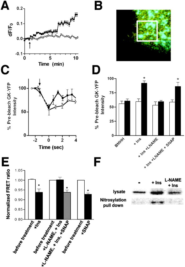
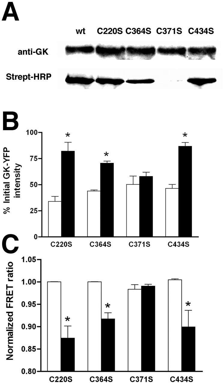
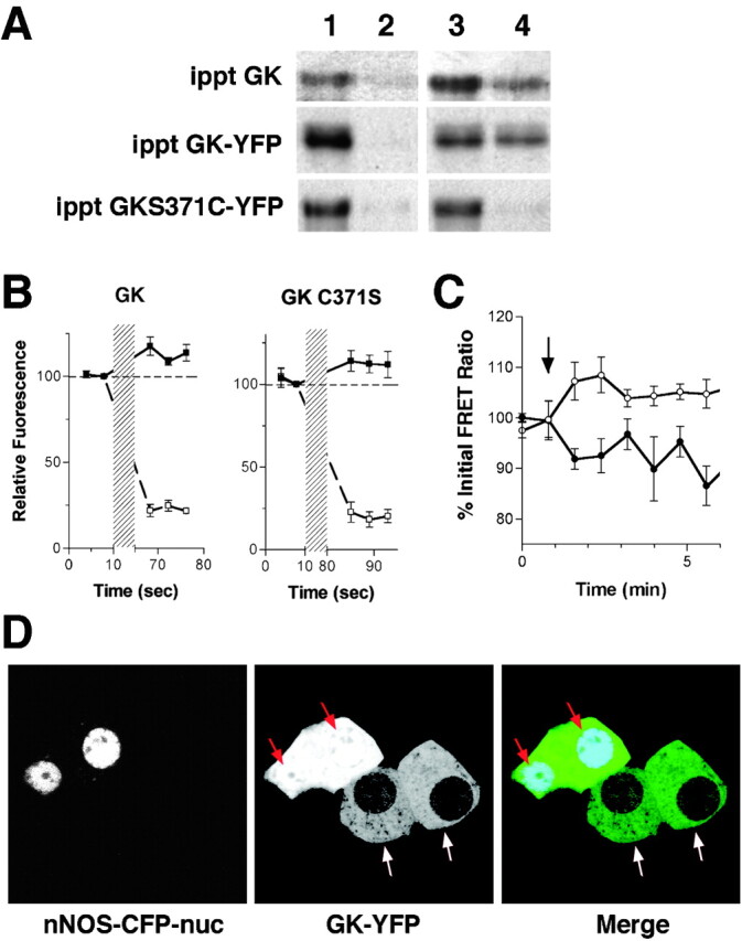
Glucokinase (GK) activity plays a key role in glucose-stimulated insulin secretion from pancreatic beta cells. Insulin regulates GK activity by modulating its association with secretory granules, although little is known about the mechanisms involved in regulating this association. Using quantitative imaging of multicolor fluorescent proteins fused to GK, we found that the dynamic association of GK with secretory granules is modulated through nitric oxide (NO). Our results in cultured beta cells show that insulin stimulates NO production and leads to S-nitrosylation of GK. Furthermore, inhibition of NO synthase (NOS) activity blocks insulin-stimulated changes in both GK association with secretory granules and GK conformation. Mutation of cysteine 371 to serine blocks S-nitrosylation of GK and causes GK to remain tightly bound to secretory granules. GK was also found to interact stably with neuronal NOS as detected by coimmunoprecipitation and fluorescence resonance energy transfer. Finally, attachment of a nuclear localization signal sequence to NOS drives GK to the nucleus in addition to its normal cytoplasmic and granule targeting. Together, these data suggest that the regulation of GK localization and activity in pancreatic beta cells is directly related to NO production and that the association of GK with secretory granules occurs through its interaction with NOS.


Figures

Figure 1.
Figure 1.
Regulation of GK by insulin requires NO. (A) βTC3 cells were starved for 4 h before loading with DAF-FM and analysis using confocal microscopy. The change in DAF intensity is represented as the change in fluorescence/initial fluorescence (dF/F0) from the average of at least 10 cells treated with 100 nM insulin (indicated by the arrow, ▪) or left untreated (○). (B) FRAP measurements were taken in cells expressing GK-YFP (yellow) and proinsulin-CFP (cyan) by selectively photobleaching YFP fluorescence in a small region (white box) of the cell containing several granules. (C) Fluorescence recovery of GK-YFP to CFP-labeled insulin granules was measured after photobleaching GK-YFP in starved cells (○) and after insulin treatment (100 nM, 5 min, ▪). The bleaching period is indicated by the arrow and broken lines. (D) FRAP measurements show fluorescence intensity of granule-associated GK-YFP immediately after photobleaching (white bars) and 2 s after photobleaching (black bars) and expressed as the percentage of prebleached fluorescence intensity. Cells were starved previously in BMHH for 3 h before insulin treatment (5 min), pretreatment with L-NAME (5 mM, 10 min) before insulin treatment (100 nM, 5 min), and after treatment with SNAP (100 μM, 1 min). Statistical significance from initial postbleach intensity (P < 0.05, t test) is denoted by an asterisk. (E) Cells expressing CFP-GK-YFP were examined for FRET by fluorescence microscopy as indicated. Statistical significance (P < 0.05 by ANOVA or t test as appropriate) is denoted by an asterisk. Cells were treated under the same conditions as in D. (F) Nitrosylated proteins were precipitated with neutravidin-agarose after biotinylation of S-nitrosylated proteins from cell lysates. Cells were treated as above where indicated. GK was detected in unreacted lysates and precipitated fractions by Western blot using an antibody to GK (Jetton and Magnuson, 1992).
Figure 2.
Figure 2.
Nitrosylation of cysteine 371 is required for GK regulation. (A) Mutant GK-YFP constructs were expressed in βTC3 cells. Nitrosylated proteins were biotinylated before immunoprecipitation of GK-YFP proteins and analysis by Western blot for GK (anti-GK) or biotinylated proteins using peroxidase-conjugated streptavidin (Strept-HRP). (B) Association of the GK-YFP mutants with secretory granules was measured using FRAP in cells expressing the indicated GK-YFP construct and proinsulin-CFP. Fluorescence intensity of granule-associated GK-YFP is expressed as the percentage of prebleached intensity immediately after bleaching (white bars) and 2 s into recovery (black bars). Statistical significance from initial postbleach intensity (P < 0.05, t test) is denoted by an asterisk. (C) Mutations were made in CFP-GK-YFP and expressed in βTC3 cells. Cells were starved for 4 h in BMHH, and the normalized FRET ratio was calculated before (white bars) and after (black bars) stimulation with 100 nM insulin (5 min). Statistical significance from pretreatment FRET ratio (P < 0.05, t test) is denoted by an asterisk.
Figure 3.
Figure 3.
Association of GK with nNOS can direct GK localization. (A) Endogenous GK, GK-YFP, and C371S GK-YFP were immunoprecipitated from cell lysates using anti-GK antibodies or anti-GFP antibodies for YFP-tagged proteins in combination with agarose-conjugated secondary antibodies. Precipitates were then heated to 37°C for 10 min in the presence of 1 mM DEANO in PBS (lanes 3 and 4) or an equivalent volume of vehicle (DMSO) in PBS alone (lanes 1 and 2). Pellets (lanes 1 and 3) and supernatants (lanes 2 and 4) were analyzed by SDS-PAGE and Western blot using anti-nNOS antibodies. (B) Cells expressing nNOS-CFP and GK-YFP or GK(C371S)-YFP were examined by two-photon microscopy before and after photobleaching with a 514 nm Ar laser (indicated by the hatched region). Average relative fluorescence (n = 6) for cellular CFP (▪) and YFP (□) intensities were plotted versus time. A dotted line was drawn as a reference to indicate prebleach intensity. (C) FRET between nNOS-CFP and either GK-YFP (•) or GK(C371S)-YFP(○) was examined in living cells by two-photon microscopy. FRET ratios were normalized to pretreatment values (100%) before averaging (n = 6) and plotted versus time. Addition of insulin (100 nM) is indicated by the arrow. (D) Cells were cotransfected with GK-YFP and nNOS-CFP-nuc and examined by confocal microscopy. In cells expressing both constructs, GK-YFP is found colocalized with nNOS in the nucleus (red arrows). GK-YFP did not localize to the nuclei of cells that were singly transfected with GK-YFP (white arrows). In the merged panel, the CFP image was colored blue and the YFP image was colored green. Colocalization is indicated by cyan (red arrows).

References

    1. Aspinwall, C.A., W.J. Qian, M.G. Roper, R.N. Kulkarni, C.R. Kahn, and R.T. Kennedy. 2000. Roles of insulin receptor substrate-1, phosphatidylinositol 3-kinase, and release of intracellular Ca2+ stores in insulin-stimulated insulin secretion in β-cells. J. Biol. Chem. 275:22331–22338. - PubMed
    1. Brenman, J.E., D.S. Chao, S.H. Gee, A.W. McGee, S.E. Craven, D.R. Santillano, Z. Wu, F. Huang, H. Xia, M.F. Peters, et al. 1996. Interaction of nitric oxide synthase with the postsynaptic density protein PSD-95 and alpha1-syntrophin mediated by PDZ domains. Cell. 84:757–767. - PubMed
    1. Fang, M., S.R. Jaffrey, A. Sawa, K. Ye, X. Luo, and S.H. Snyder. 2000. Dexras1: a G protein specifically coupled to neuronal nitric oxide synthase via CAPON. Neuron. 28:183–193. - PubMed
    1. Henningsson, R., A. Salehi, and I. Lundquist. 2002. Role of nitric oxide synthase isoforms in glucose-stimulated insulin release. Am. J. Physiol. Cell Physiol. 283:C296–C304. - PubMed
    1. Jaffrey, S.R., H. Erdjument-Bromage, C.D. Ferris, P. Tempst, and S.H. Snyder. 2001. Protein S-nitrosylation: a physiological signal for neuronal nitric oxide. Nat. Cell Biol. 3:193–197. - PubMed

Publication types

MeSH terms