. 2003 Apr 28;161(2):281-94.
doi: 10.1083/jcb.200208092. Epub 2003 Apr 21.

The small molecule Hesperadin reveals a role for Aurora B in correcting kinetochore-microtubule attachment and in maintaining the spindle assembly checkpoint

Affiliations

The small molecule Hesperadin reveals a role for Aurora B in correcting kinetochore-microtubule attachment and in maintaining the spindle assembly checkpoint

Silke Hauf et al. J Cell Biol. .

Abstract

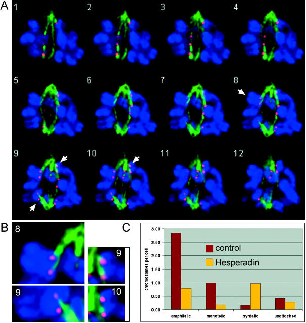
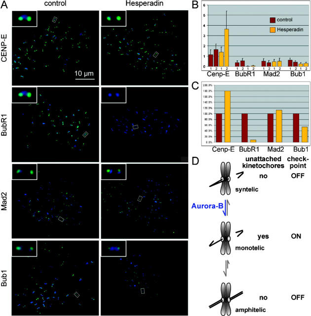
The proper segregation of sister chromatids in mitosis depends on bipolar attachment of all chromosomes to the mitotic spindle. We have identified the small molecule Hesperadin as an inhibitor of chromosome alignment and segregation. Our data imply that Hesperadin causes this phenotype by inhibiting the function of the mitotic kinase Aurora B. Mammalian cells treated with Hesperadin enter anaphase in the presence of numerous monooriented chromosomes, many of which may have both sister kinetochores attached to one spindle pole (syntelic attachment). Hesperadin also causes cells arrested by taxol or monastrol to enter anaphase within <1 h, whereas cells in nocodazole stay arrested for 3-5 h. Together, our data suggest that Aurora B is required to generate unattached kinetochores on monooriented chromosomes, which in turn could promote bipolar attachment as well as maintain checkpoint signaling.


Figures

Figure 1.
Figure 1.
Hesperadin causes polyploidy in HeLa cells. (A) Chemical structure of Hesperadin. (B) Hesperadin (50 nM) was added to logarithmically growing HeLa cells (day 0). At the indicated time points, the DNA content was determined by flow cytometry, and phase contrast micrographs were taken. From day 4 on, the DNA content could not be measured by flow cytometry (n.a., not applicable). (C) HeLa cells were synchronized by double thymidine treatment and released either into 100 nM Hesperadin (right) or a corresponding concentration (0.01%) of the solvent DMSO (left). Cells were harvested at the indicated time points after release, and the DNA content was analyzed by flow cytometry. 330 nM nocodazole was added to one sample of each series, and these cells were harvested at 14.5 h after release from thymidine. log, logarithmically growing untreated HeLa cells. (D) Samples from the same experiment as in C were analyzed by SDS-PAGE and immunoblotting.
Figure 2.
Figure 2.
Hesperadin inhibits histone H3 phosphorylation and causes midspindle defects. (A) Samples from the same experiment as in Fig. 1 (C and D) were analyzed by SDS-PAGE and immunoblotting with anti–phospho-histone H3 antibody. (B) HeLa cells were treated with Hesperadin or DMSO (control) for 16 h and immunostained with anti–phospho-histone H3. DNA was stained with DAPI. Chromosomes that have not aligned at the metaphase plate are indicated by arrowheads. (C) HeLa cells were treated as in B. Cells were costained with α-tubulin and either CYK-4 or MKLP-1 antibodies. DNA was stained with DAPI. In Hesperadin-treated cells, chromatin is not segregated properly in anaphase, indicated by arrows. (D) Cdk1 or human Aurora B were immunoprecipitated from mitotic HeLa cell extracts, and in vitro kinase activity was determined using histone H1 or histone H3, respectively, as a substrate. Hesperadin was added to the kinase reactions at the indicated concentrations. As a control, 0.1% DMSO was added (corresponding to the DMSO concentration in the sample containing 5,000 nM Hesperadin). Further controls included a kinase reaction without substrate as well as mock immunoprecipitates by unspecific antibodies.
Figure 3.
Figure 3.
Aurora B RNAi in human cells induces a similar phenotype as Hesperadin. (A) HeLa cells were transfected with Aurora B–targeting siRNA duplex (RNAi AurB) or with H2O replacing the siRNA (control). After 50 h, cells were lysed in sample buffer. Samples were resolved by SDS-PAGE and immunoblotted. (B) Cells treated as in A were processed for immunofluorescence 48 h after transfection. Cells were costained for Aurora B (in green) and phospho-histone H3 (in red). DNA was stained with DAPI (left panel, right panel in blue, also in C and D). Chromosomes that have not aligned to the metaphase plate are indicated by arrowheads. (C and D) Cells treated as in A were processed for immunofluorescence 72 h after transfection. Cells were stained for α-tubulin (in green) and Survivin (C, in red). Cells treated with Aurora B siRNA duplex did not form a proper midspindle and showed segregation defects, indicated by an arrow.
Figure 4.
Figure 4.
Hesperadin-treated HeLa cells show alignment and segregation defects, but sister chromatid separation is intact. HeLa cells were left untreated or were treated with 50 nM Hesperadin for different periods of time before harvesting by mitotic shake off, followed by chromosome spreading and Giemsa staining. (a) Normal metaphase plate. Inset, normal degree of sister chromatid resolution. Compare the gap between sisters (arrow) in a and c. (b) Normal onset of anaphase. (c) Typical appearance of a late prometaphase in Hesperadin-treated cultures. Defects in alignment and sister chromatid resolution (arrow) are apparent. Some chromosomes are bent at the centromeric region (inset, examples marked by squares), implying microtubule attachment. (d–f) Typical appearance of early anaphases in Hesperadin-treated cells. Chromosomes often seem to be segregated to opposite poles, but many sister chromatids stay in close proximity (double arrows). One example of sister chromatids being pulled to opposite poles is marked by arrowheads. (g and h) Typical appearance of chromatin decondensation in Hesperadin-treated cells. (i) Typical interphase in Hesperadin-treated cultures.
Figure 5.
Figure 5.
Aurora B function is required at the time of microtubule attachment to kinetochores. (A and C) HeLa cells were arrested in nocodazole. Hesperadin or the solvent DMSO were added shortly before release from nocodazole (outlined in A). Chromosome spreads were performed with the arrested cells after addition of Hesperadin or DMSO (noc-arrest), directly after washing out nocodazole (0'), and at different time points after release from nocodazole (15–150'). Representative images are shown in C. For a quantification, see Fig. S2 (available at http://www.jcb.org/cgi/content/full/jcb.200208092/DC1). (B and D) HeLa cells were arrested in nocodazole in the presence of Hesperadin or the solvent DMSO. Hesperadin and DMSO were washed out before releasing cells from nocodazole (outlined in B). Chromosome spreads were performed with the arrested cells after Hesperadin or DMSO washout (noc-arrest), directly after washing out nocodazole (0'), and at different time points after release from nocodazole (15–150'). Representative images are shown in D. For a quantification, see Fig. S2.
Figure 6.
Figure 6.
Monoorientation is not corrected in Hesperadin-treated PtK1 cells before anaphase. Live cell imaging of a PtK1 cell treated with 500 nM Hesperadin. Selected stills of a time-lapse movie (Video 2, available at http://www.jcb.org/cgi/content/full/jcb.200208092/DC1), elapsed time from NEB in h:min is shown in the upper left. The approximate positions of the spindle poles are marked by yellow stars. Red dots indicate the centromeric region of two different chromosomes. After NEB (0:00), some chromosomes align to the metaphase plate, but the majority of chromosomes are monooriented (0:17–0:44). Monooriented chromosomes move on the spindle (compare distance between red dots and yellow stars), but they fail to congress. Upon the onset of anaphase, sister chromatids of bioriented chromosomes are pulled to opposite poles (example marked by red arrowheads), whereas both sisters of the monooriented chromosomes move to the same pole.
Figure 7.
Figure 7.
Hesperadin-treated PtK cells show a higher frequency of syntelic attachments during prometaphase than control-treated cells. (A and B) PtK1 cell treated with 500 nM Hesperadin for 3 h. Deconvolved images taken at 0.2-μm Z-interval. α-Tubulin (green), CREST (red), and DNA staining (blue). Chromosomes that are attached in a syntelic manner are marked by arrows and are enlarged in B. (C) PtK2 cells were treated with 500 nM Hesperadin or 0.1% DMSO (control) for 3 h. Deconvolution microscopy was performed as in A, and the type of attachment was determined for as many chromosomes as possible (control: 26 cells, 115 chromosomes; Hesperadin: 29 cells, 64 chromosomes; see Fig. S3, available at http://www.jcb.org/cgi/content/full/jcb.200208092/DC1). The average number of chromosomes per cell exhibiting a certain type of attachment is shown.
Figure 8.
Figure 8.
Hesperadin quickly overrides the mitotic arrest induced by taxol or monastrol. (A, B, and C) HeLa cells were arrested in nocodazole (330 nM), taxol (10 μM), or monastrol (100 μM). Hesperadin (100 nM) or the solvent DMSO was added, and cells were followed by chromosome spreading (A and B) or immunofluorescence microscopy (C). For immunofluorescence microscopy (C), cells were stained with α-tubulin (left), DNA was counterstained with DAPI (right). (D) Percentage of cells arrested in mitosis in the presence of the different drugs is shown as a function of time. Numbers were obtained from the samples shown in A–C. (E) PtK1 cells were treated with monastrol, and mitotic entry was followed by differential interference contrast microscopy. Selected stills of a time-lapse movie are shown, elapsed time in h:min is shown in the lower right. 500 nM Hesperadin was added at time point 1:10 when the typical monoastral spindle had formed, and mitotic exit was followed. All sister chromatids move into the single polar region.
Figure 9.
Figure 9.
BubR1 localization to kinetochores is abolished in HeLa cells treated with nocodazole and Hesperadin. (A) HeLa cells were arrested in mitosis with 10 μM nocodazole and then additionally treated with 100 nM Hesperadin or the solvent DMSO for 2 h, harvested by mitotic shake off, cytospun on slides, and processed for immunofluorescence with the indicated antibodies (in green). Kinetochores were labeled with CREST serum (in blue). The insets show magnifications of the kinetochore pairs marked by white rectangles. (B) Data, as in A, were quantified. For each cell, the average integrated intensity for the checkpoint protein was related to the average integrated intensity of the CREST signal. Bars show the average of the ratios obtained from 10 cells. The reduction of BubR1 signal intensity was significant in both independent experiments (1 and 2) (P < 0.0001, t test), whereas the reduction of Bub1 signal intensity was only significant in experiment 1 (experiment 1, P < 0.0001; experiment 2, P = 0.059). (C) Average of the two independent experiments shown in B, with the control data set to 100%. (D) Model for Aurora B function.

References

    1. Abruzzi, K.C., M. Magendantz, and F. Solomon. 2002. An α-tubulin mutant demonstrates distinguishable functions among the spindle assembly checkpoint genes in Saccharomyces cerevisiae. Genetics. 161:983–994. - PMC - PubMed
    1. Adams, R.R., M. Carmena, and W.C. Earnshaw. 2001. a. Chromosomal passengers and the (aurora) ABCs of mitosis. Trends Cell Biol. 11:49–54. - PubMed
    1. Adams, R.R., H. Maiato, W.C. Earnshaw, and M. Carmena. 2001. b. Essential roles of Drosophila inner centromere protein (INCENP) and aurora B in histone H3 phosphorylation, metaphase chromosome alignment, kinetochore disjunction, and chromosome segregation. J. Cell Biol. 153:865–880. - PMC - PubMed
    1. Ault, J.G., and C.L. Rieder. 1992. Chromosome mal-orientation and reorientation during mitosis. Cell Motil. Cytoskeleton. 22:155–159. - PubMed
    1. Biggins, S., and A.W. Murray. 2001. The budding yeast protein kinase Ipl1/Aurora allows the absence of tension to activate the spindle checkpoint. Genes Dev. 15:3118–3129. - PMC - PubMed

Publication types

MeSH terms