. 2003 May;109(1):15-23.
doi: 10.1046/j.1365-2567.2003.01598.x.

Widespread inosine-containing mRNA in lymphocytes regulated by ADAR1 in response to inflammation

Affiliations

Widespread inosine-containing mRNA in lymphocytes regulated by ADAR1 in response to inflammation

Jing-Hua Yang et al. Immunology. 2003 May.

Abstract

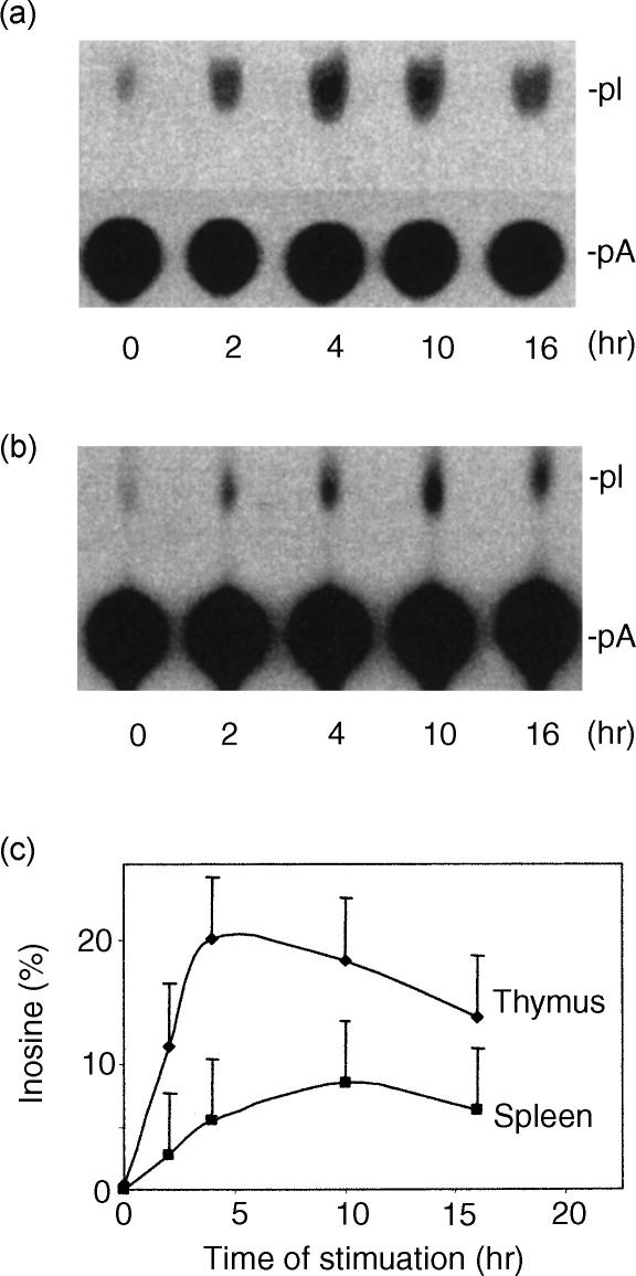
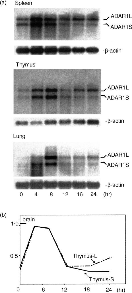
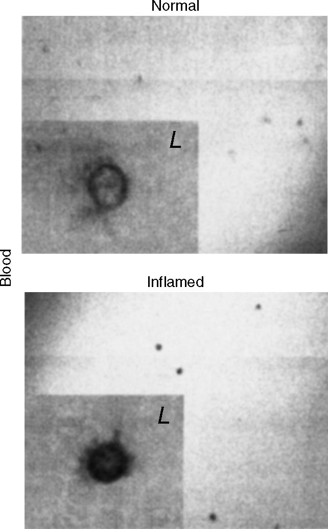
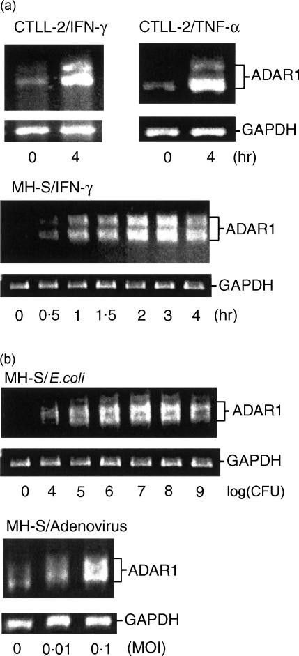
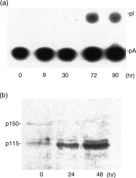
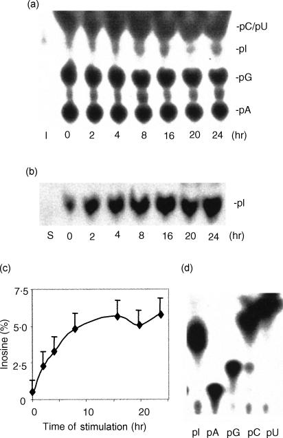
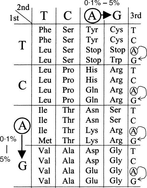
Adenosine-to-inosine (A-to-I) RNA editing is a post-transcriptional modification of pre-mRNA catalysed by an RNA-specific adenosine deaminase (ADAR). A-to-I RNA editing has been previously reported in the pre-mRNAs of brain glutamate and serotonin receptors and in lung tissue during inflammation. Here we report that systemic inflammation markedly induces inosine-containing mRNA to approximately 5% of adenosine in total mRNA. Induction was the result of up-regulation of A-to-I RNA editing as both dsRNA editing activity and ADAR1 expression were increased in the spleen, thymus and peripheral lymphocytes from endotoxin-treated mice. Up-regulation of ADAR1 was confirmed in vitro in T lymphocytes and macrophages stimulated with a variety of inflammatory mediators including tumour necrosis factor-alpha and interferon-gamma. A late induction of RNA editing was detected in concanavalin A-activated splenocytes stimulated with interleukin-2 in vitro. Taken together, these data suggest that a large number of inosine-containing mRNAs are produced during acute inflammation via up-regulation of ADAR1-mediated RNA editing. These events may affect the inflammatory and immune response through modulation of protein production.


Figures

Figure 1
Figure 1
Up-regulation of A-to-I RNA editing in mouse thymus (a) and spleen (b) during acute systemic inflammation. Spleens and thymuses were harvested (n = 6) at the indicated time-points after endotoxin-challenge and whole cell extracts were examined for editing activity. Editing activity was determined by measuring A-to-I conversion of dsRNA on TLC. The percentage of inosines was estimated in comparison with adenosines and plotted against the duration of endotoxin stimulation (c). pI, 5′-monophosphate inosine; pA, 5′-monophosphate adenosine. The ratio between pI and pA represents the percentage of adenosine that is converted to inosine.
Figure 2
Figure 2
Up-regulation of ADAR1 expression in acute systemic inflammation assayed by Northern blotting. (a) Mouse spleen, thymus, and lung were harvested (n = 6) at different time-points as indicated after endotoxin-challenge. Four micrograms of mRNA were isolated and analysed by Northern blotting. ADAR1 mRNA was detected using synthesized oligo complementary ADAR1 sequences from 1305 to 1265 and 3004 to 2996 (AF291050). Two transcripts, ADAR1L and ADAR1S, were observed in all organs. (b) ADAR1 mRNA was quantified by PhospherImager and normalized to the internal control β-actin. The ratios between ADAR1L and β-actin and of ADAR1S and β-actin were calculated to compare their relative expression. Note that the calculated ratios were similar prior to and early after induction of inflammation. ADAR1L became dominant later (>12 hr).
Figure 3
Figure 3
Cellular localization of ADAR1 in peripheral blood during acute systemic inflammation by in situ hybridization. In situ hybridization was applied on mouse peripheral blood smear for the detection of ADAR1 mRNA using digoxigenin-labelled antisense ADAR1 RNA probes (1305–1265 and 3014–2972, AF291050). Positive signals were identified in lymphocytes (L). Similar observations were uniformly seen in all animals (n = 6).
Figure 4
Figure 4
Induction of ADAR1 by inflammatory mediators (a) and pathogens (b) in vitro. (a) Cytotoxic T cells (CTLL-2) and macrophages (MH-S) were stimulated with IFN-γ (1000 U/ml) or TNF-α (10 ng/ml). In both cell types RT-PCR analysis indicated a marked increase in ADAR1 after 4 hr of stimulation. (b) Macrophages (5 × 10⁶ MH-S cells) were stimulated with live E. coli (104−109 CFU) or adenovirus (0·01 and 0·1 MOI) for 2 hr. ADAR1 was up-regulated by both pathogens. Each gel represents a single experiment. Similar observations were uniformly seen in all experiments (n = 3). The primers for RT-PCR cover exons 5–8. Two RT-PCR products were seen in all cases because of alternative splicing in exon 7 (AF291050 and AF291876).
Figure 5
Figure 5
Late induction of RNA editing in activated splenic cells. (a) Splenic cells were harvested from sham mice (n = 6) and activated with Con A (2 μg/ml) and IL-2 (2 μg/ml) for the times indicated. Whole cell extracts were prepared for editing activity. (b) Splenic cells were harvested from sham mice (n = 6) and activated with Con A (2 µg/ml) for the times indicated. Whole cell extracts were prepared for Western blotting as described above. Note that induction of editing was observed at 30 hr in correlation with cell proliferation. On the Western blots, two ADAR1 variants (p150 and p115) were detected. Induction of the short form corresponded with the increased RNA editing activity.
Figure 6
Figure 6
Induction of inosines in thymic mRNA during acute systemic inflammation. (a) The first TLC assay. Inosine-containing mRNA was analysed in thymuses from endotoxin-stimulated mice (n = 4) at 0–24 hr. Total poly(A)+ RNA (tRNA and rRNA were not visible when monitored on denature agarose gels) from each time-point was isolated and a synthetic RNA was made by in vitro transcription using a plasmid with T7 promoter. Poly(A)+ RNA was digested with RNase T2, labelled with polynucleotide kinase and digested again with RNase P1. The inosine-labelled mRNA was significantly increased. pA, pG, pI, pC and pU are 5′-monophosphate nucleotides; I, the standard 5′-[32P]monophosphate inosine. (b) The second TLC assay. After the first TLC, inosine and adenosine were recovered and analysed on a new TLC. In agreement with the first TLC, inosine-labelled mRNA was significantly increased. S, synthetic RNA. (c) Quantification of the increased level of inosine-containing mRNA. Per cent of inosine was calculated using radioactive counting or an NIH Image software. Results were plotted against the duration of stimulation. Note the increase from a baseline of approximately 0·5–5% at 24 hr. (d) Labelling efficiency of 3′-monophosphate nucleotides. PNK equally labelled all 3′-monophosphate nucleotides (pA, pG, pC, pU and pI).
Figure 7
Figure 7
Codon switch by A-to-I RNA editing. Editing in the coding region of mRNA could switch the codon identity. The arrows indicate <0·1% codon switch in normal conditions and 5% during acute inflammation. If editing occurs at the first or second codon positions, it switches the encoded amino acids. Editing at the third codon position or outside of the coding region of inosine-containing mRNA is termed ‘silent’ editing.

Similar articles

Cited by

References

    1. Yang JH, Sklar P, Axel R, Maniatis T. Editing of glutamate receptor subunit B pre-mRNA in vitro by site-specific deamination of adenosine. Nature. 1995;374:77–81. - PubMed
    1. Melcher T, Maas S, Higuchi M, Keller W, Seeburg PH. Editing of alpha-amino-3-hydroxy-5-methylisoxazole-4-propionic acid receptor-B pre-mRNA in vitro reveals site-selective adenosine to inosine conversion. J Biol Chem. 1995;270:8566–70. - PubMed
    1. Rueter SM, Burns CM, Coode SA, Mookherjee P, Emeson RB. Glutamate receptor RNA editing in vitro by enzymatic conversion of adenosine to inosine. Science. 1995;267:1491–4. - PubMed
    1. Kim U, Wang Y, Sanford T, Zeng Y, Nishikura K. Molecular cloning of cDNA for double-stranded RNA adenosine deaminase, a candidate enzyme for nuclear RNA editing. Proc Natl Acad Sci USA. 1994;91:11457–61. - PMC - PubMed
    1. Melcher T, Maas S, Herb A, Sprengel R, Seeburg PH, Higuchi M. A mammalian RNA editing enzyme. Nature. 1996;379:460–4. - PubMed

Publication types

MeSH terms

LinkOut - more resources