. 2003 May 1;31(9):2279-88.
doi: 10.1093/nar/gkg347.

Molecular determinants of the cell-cycle regulated Mcm1p-Fkh2p transcription factor complex

Affiliations

Molecular determinants of the cell-cycle regulated Mcm1p-Fkh2p transcription factor complex

Joanna Boros et al. Nucleic Acids Res. .

Abstract

The MADS-box transcription factor Mcm1p and forkhead (FKH) transcription factor Fkh2p act in a DNA-bound complex to regulate cell-cycle dependent expression of the CLB2 cluster in Saccharomyces cerevisiae. Binding of Fkh2p requires prior binding by Mcm1p. Here we have investigated the molecular determinants governing the formation of the Mcm1p- Fkh2p complex. Fkh2p exhibits cooperativity in complex formation with Mcm1p and we have mapped a small region of Fkh2p located immediately upstream of the FKH DNA binding domain that is required for this cooperativity. This region is lacking in the related protein Fkh1p that cannot form ternary complexes with Mcm1p. A second region is identified that inhibits Mcm1p-independent DNA binding by Fkh2p. The spacing between the Mcm1p and Fkh2p binding sites is also a critical determinant for complex formation. We also show that Fkh2p can form ternary complexes with the human counterpart of Mcm1p, serum response factor (SRF). Mutations at analogous positions in Mcm1p, which are known to affect SRF interaction with its partner protein Elk-1, abrogate complex formation with Fkh2p, demonstrating evolutionary conservation of coregulatory protein binding surfaces. Our data therefore provide molecular insights into the mechanisms of Mcm1p- Fkh2p complex formation and more generally aid our understanding of MADS-box protein function.


Figures
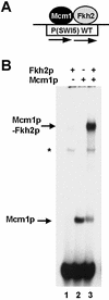
Figure 1
Figure 1
Fkh2p requires Mcm1p to bind the SWI5 promoter. (A) Schematic of the ternary Mcm1p–Fkh2p complex that forms on the composite CArG–Fkh binding motif in the SWI5 promoter. (B) Gel retardation analysis of in vitro translated Mcm1(1–98) and full-length Fkh2p on the CArG–fkh motif from the SWI5 promoter. The addition of Fkh2p and Mcm1p, and the locations of the DNA-bound Mcm1p and Mcm1p–Fkh2p complexes are indicated. The asterisk represents a non-specific band arising from the reticulocyte lysates.
Figure 2
Figure 2
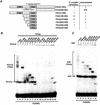
Mapping the ternary complex determinants in Fkh2p. (A) A schematic of the truncated Fkh2p constructs generated for the mapping analysis. The locations of the FHA and FKH domains are indicated. The minimal region required for ternary complex formation and the location of the autoinhibitory domain are indicated. The dotted line indicates that the C-terminal end of the autoinhibitory motif is unknown. The ability of each Fkh2p construct to form ternary complexes and exhibit autoinhibitory DNA binding properties is indicated. (B and C) Gel retardation analysis of the indicated Fkh2p proteins in the presence or absence of Mcm1(1–98) (B) or in the presence of SRF(132–222) on the P(SWI5) site. Asterisks indicate Fkh2p truncations that bind DNA in the absence of Mcm1p.
Figure 3
Figure 3
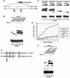
Fine mapping of the Fkh2p ‘cooperativity domain’. (A) A schematic of the truncated Fkh2(292–458) protein. The amino acid sequence of the sequence immediately preceding the FKH domain of Fkh2p and the analogous region in Fkh1p are shown. The amino acids at the truncation endpoints are highlighted. (B) Gel retardation analysis of the indicated Fkh2p truncated proteins in the absence (lanes 1–4) or presence (lanes 5–8) of Mcm1(1–96) on the P(SWI5) site. (C) Gel retardation analysis of increasing amounts of the truncated Fkh2p truncated proteins in the presence (lanes 1–10) or absence (lanes 11–20) of Mcm1(1–96). Relative molar amounts of Fkh2p were 1 (lanes 1, 6, 11 and 16); 3 (lanes 2, 7, 12, 17); 5 (lanes 3, 8, 13, 18); 10 (lanes 4, 9, 14, 19); 20 (lanes 5, 10, 15, 20). (D) Quantification of the data in (C). The binding observed with the highest concentrations of Fkh2p used is not shown as the binding in lane 5 is beyond the linear range. (E) Alignment of amino acids 312–333 of Fkh2p with the B-box region (amino acids 150–168) of Elk-1. Amino acids in Elk-1 that affect interactions with SRF by >50% (18) are shaded. These are conserved in Fkh2p. (F) Gel retardation analysis of the indicated Fkh2p mutant proteins in the absence (lanes 1–3) or presence (lanes 4–6) of Mcm1(1–96) on the P(SWI5) site.
Figure 4
Figure 4
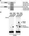
Fkh1p does not bind cooperatively with Mcm1p. (A) Schematic of full-length and truncated versions of Fkh1p and Fkh2p. (B) Gel retardation analysis of the indicated Fkh1p and Fkh2p truncated proteins in the absence (lanes 1–4) or presence (lanes 5–8) of Mcm1(1–98) on the P(SW15) site. The locations of complexes corresponding to Mcm1p alone, Fkh1p/Fkh2p alone or Mcm1p–Fkh2p complexes are indicated. The asterisk indicates the location of a band corresponding to a weak ternary complex.
Figure 5
Figure 5
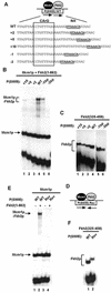
Influence of binding site spacing on Mcm1p–Fkh2p complex formation. (A) Schematic of the Mcm1p–Fkh2p ternary complex bound to the P(SWI5) site. The sequence of the wild-type site and ‘spacer’ mutants are shown below. The fkh binding motif is shown in bold and underlined and the CArG box Mcm1p binding motif is boxed. (B and C) Gel retardation analysis of Fkh2(1–862) (B) and Fkh2(325–458) (C) in the presence (B) or absence (C) of Mcm1(1–96) bound to the indicated wild-type (WT) and ‘spacer’ mutant binding sites. (D) Schematic of the Mcm1p–Fkh2p complex and the P(SW15)Rev binding site. The arrows represent the relative orientation of the Mcm1p and Fkh2p binding sites. (E and F) Gel retardation analysis of Fkh2(1–862) (E) and Fkh2(325–458) (F) in the presence (E) and absence (F) of Mcm1p on the WT and Rev versions of the P(SWI5) site. The locations of Mcm1p, Fkh2p and Mcm1p–Fkh2p complexes are indicated in each panel.
Figure 6
Figure 6
Identification of the Fkh2p binding surface on Mcm1p. (A) Alignment of the Elk-1 binding regions in SRF with the equivalent region in Mcm1p. Residues mutated in this study are highlighted. Dots between the two sequences indicate identical amino acids. Residues comprising secondary structural elements are bracketed. (B) A structural representation of Mcm1p bound to DNA (monomers coloured in yellow and orange) (19). The residues mutated in this study are shown in blue. (C and D) Gel retardation analysis of the indicated Mcm1(1–98) mutants in the absence (C and D, lanes 1–5) or the presence of either Fkh2(1–862) (C, lanes 6–10) or Fkh2(254–458) (D, lanes 6–10) on the P(SWI5) site. The locations of complexes corresponding to Mcm1p alone or Mcm1p–Fkh2p are indicated. The amount of binary Mcm1p complex was normalised to give equivalent binding. (E) A GST pulldown experiment of GST–Fkh2(254–458) and the indicated in vitro translated Mcm1(1–98) mutants. Ten percent of input protein is shown. Quantification of the data normalised to input protein is shown below and compared with wild-type Mcm1p (taken as ‘100’). The average of three independent experiments is shown.
Figure 7
Figure 7
The role of Mcm1p–Fkh2p interactions in vivo. (A) The mcm1Δ mutant strain, YY2052, was transformed with plasmids encoding wild-type or V69E version of Mcm1(1–98) and plated on SD or FOA plates (to remove the plasmid-borne mcm1 allele). (B) The fkh2Δndd1Δ double mutant (AP183) was transformed with pMW20-NDD1 and either the pGBKT7 vector (control), pGBKT7–Fkh2(1–458) or pGBKT7–Fkh2(1–458)(Y315E) plasmids. Neat and 10-fold serial dilutions of transformants were spotted onto SD and FOA plates. Plates were incubated at 30°C for 2–5 days.
Figure 8
Figure 8
Binding sites for the Mcm1p–Fkh2p complex. Potential binding motifs are shown for the Mcm1p–Fkh2p complex located within the promoters of genes revealed to be direct targets of the Mcm1p–Fkh2p–Ndd1p complex by chromatin IP analysis (37). The Mcm1p and Fkh2p binding sites (boxed and shaded) and spacer region are shown. A conserved dinucleotide motif in the spacer is underlined.

Similar articles

Cited by

References

    1. Dolan J.W. and Fields,S. (1991) Cell-type-specific transcription in yeast. Biochim. Biophys. Acta, 1088, 155–169. - PubMed
    1. Shore P. and Sharrocks,A.D. (1995) The MADS-box family of transcription factors. Eur. J. Biochem., 229, 1–13. - PubMed
    1. McInerny C.J., Partridge,J.F., Mikesell,G.E., Creemer,D.P. and Breeden,L.L. (1997) A novel Mcm1-dependent element in the SWI4, CLN3, CDC6, and CDC47 promoters activates M/G1-specific transcription. Genes Dev., 11, 1277–1288. - PubMed
    1. Althoefer H., Schleiffer,A., Wassmann,K., Nordheim,A. and Ammerer,G. (1995) Mcm1 is required to coordinate G2-specific transcription in Saccharomyces cerevisiae. Mol. Cell. Biol., 15, 5917–5928. - PMC - PubMed
    1. Breeden L.L. (2000) Cyclin transcription: timing is everything. Curr. Biol., 10, R586–588. - PubMed

Publication types

MeSH terms