. 2003 May 1;31(9):2323-32.
doi: 10.1093/nar/gkg339.

Steady-state and time-resolved fluorescence studies indicate an unusual conformation of 2-aminopurine within ATAT and TATA duplex DNA sequences

Affiliations

Steady-state and time-resolved fluorescence studies indicate an unusual conformation of 2-aminopurine within ATAT and TATA duplex DNA sequences

Priyamvada Rai et al. Nucleic Acids Res. .

Abstract

2-Aminopurine (2-AP), a fluorescent analog of adenine, has been widely used as a probe for local DNA conformation, since excitation and emission characteristics and fluorescence lifetimes of 2-AP vary in a sequence-dependent manner within DNA. Using steady-state and time-resolved fluorescence techniques, we report that 2-AP appears to be unusually stacked in the internal positions of ATAT and TATA in duplex DNA. The excitation wavelength maxima for 2-AP within these contexts were red shifted, indicating reduced solvent exposure for the fluorophore. Furthermore, in these contexts, 2-AP fluorescence was resistant to acrylamide-dependent collisional quenching, suggesting that the fluorophore is protected by its stacked position within the duplex. This conclusion was further reinforced by the presence of a secondary peak at 275 nm in the fluorescence excitation spectra that is indicative of efficient excitation energy transfer from nearby non-fluorescent DNA bases. Fluorescence anisotropy decay and internal angular 'wobbling' motion measurements of 2-AP within these alternating AT contexts were also consistent with the fluorophore being highly constrained and immobile within the base stack. When these fluorescence characteristics are compared with those of 2-AP within other duplex DNA sequence contexts, they are unique.


Figures
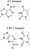
Figure 1
Figure 1
Schematic of Watson–Crick adenine-thymine (A-T) and 2-aminopurine-thymine (2-AP-T) base pairs.
Figure 2
Figure 2
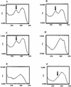
Representative fluorescence excitation spectra. Excitation profiles were taken at 30°C and automatically printed by the Hitachi-F2000 fluorometer. They were then scanned and traced over using the ‘autotrace’ function of Canvas 7.0 to improve the print quality of the plotter-generated spectra. The wavelength (in nm) is on the x-axis and excitation intensity (in absorbance units) is on the y-axis and is denoted by I. Excitation energy transfer peaks are indicated with arrows. Spectra are shown for: (A) ds.P7; (B) ds.P27; (C) ds.P26; (D) ds.P12; (E) ss.P8; (F) ds.P7_eh.
Figure 3
Figure 3
Effect of acrylamide upon the fluorescence of 2-AP in different contexts. Acrylamide was added at 30°C to give a final concentration of 50 mM per increment as described in Materials and Methods. (A) Stern–Volmer plots for single-stranded (ss.P26) and duplexed (ds.P26) oligonucleotides. (B) Bimolecular quenching rate constants obtained from analysis of Stern–Volmer plots according to equations 3 and 5 (Materials and Methods).
Figure 3
Figure 3
Effect of acrylamide upon the fluorescence of 2-AP in different contexts. Acrylamide was added at 30°C to give a final concentration of 50 mM per increment as described in Materials and Methods. (A) Stern–Volmer plots for single-stranded (ss.P26) and duplexed (ds.P26) oligonucleotides. (B) Bimolecular quenching rate constants obtained from analysis of Stern–Volmer plots according to equations 3 and 5 (Materials and Methods).
Figure 4
Figure 4
Summary of sequence-dependent anomalies in 2-AP conformation within the ATAT-containing and TATA-containing duplexes. The positions of the various 2-AP substitutions studied within the two duplexes are underlined. The positions at which 2-AP exhibited red-shifted excitation maxima and energy transfer peaks at 275 nm as well as low rotational motion are shown (outlined) within the ATAT- and TATA-containing duplexes. In each instance, 2-AP falls within a staggered duplexed TAT context.

References

    1. Sowers L.C., Fazakerly,G.V., Eritja,R., Kaplan,B.E. and Goodman,M.F. (1986) Base pairing and mutagenesis: observation of a protonated base pair between 2-aminopurine and cytosine in an oligonucleotide by proton NMR. Proc. Natl Acad. Sci. USA, 83, 5434–5438. - PMC - PubMed
    1. Eritja R., Kaplan,B.E., Mhaskar,D., Sowers,L.C., Petruska,J. and Goodman,M.F. (1986) Synthesis and properties of defined DNA oligomers containing base mispairs involving 2-aminopurine. Nucleic Acids Res., 14, 5869–5884. - PMC - PubMed
    1. Stivers J.T. (1998) 2-Aminopurine fluorescence studies of base stacking interactions at abasic sites in DNA: metal-ion and base sequence effects. Nucleic Acids Res., 26, 3837–3844. - PMC - PubMed
    1. Rachofsky E.L., Osman,R. and Ross,J.B.A. (2001) Probing structure and dynamics of DNA with 2-aminopurine: effects of local environment on fluorescence. Biochemistry, 40, 946–956. - PubMed
    1. Rachofsky E.L., Seibert,E., Stivers,J.T., Osman,R. and Ross,J.B.A. (2001) Conformation and dynamics of abasic sites in DNA investigated by time-resolved fluorescence of 2-aminopurine. Biochemistry, 40, 957–967. - PubMed

Publication types