. 2003 May 1;31(9):2369-80.
doi: 10.1093/nar/gkg346.

HMG box transcription factor TCF-4's interaction with CtBP1 controls the expression of the Wnt target Axin2/Conductin in human embryonic kidney cells

Affiliations

HMG box transcription factor TCF-4's interaction with CtBP1 controls the expression of the Wnt target Axin2/Conductin in human embryonic kidney cells

Tomas Valenta et al. Nucleic Acids Res. .

Abstract

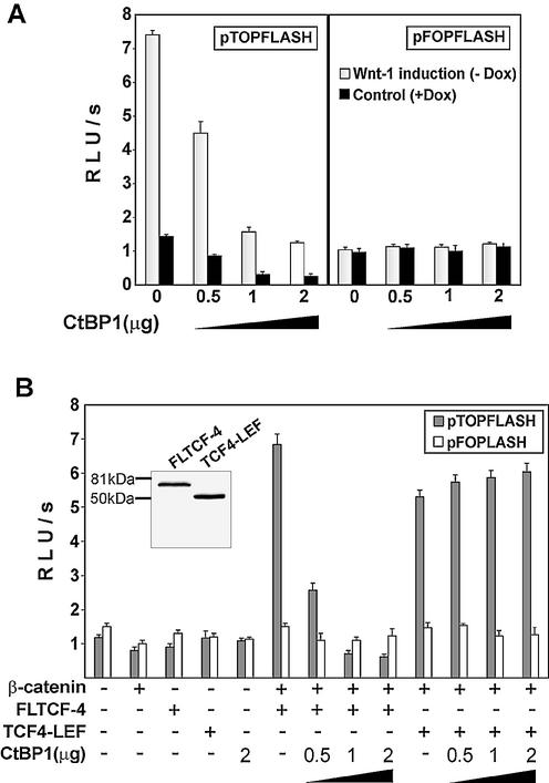
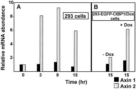
Members of the Tcf/Lef family of the HMG box transcription factors are nuclear effectors of the Wnt signal transduction pathway. Upon Wnt signaling, TCF/LEF proteins interact with beta-catenin and activate transcription of target genes, while, in the absence of the Wnt signal, TCFs function as transcriptional repressors. All vertebrate Tcf/Lef transcription factors associate with TLE/Groucho-related co-repressors, and here we provide evidence for an interaction between the C-terminus of the TCF-4 HMG box protein and the C-terminal binding protein 1 (CtBP1) transcriptional co-repressor. Using Wnt-1-stimulated human embryonic kidney 293 cells, we show that CtBP1 represses the transcriptional activity of a Tcf/beta-catenin-dependent synthetic promoter and, furthermore, decreases the expression of the endogenous Wnt target, Axin2/Conductin. The CtBP1-mediated repression was alleviated by trichostatin A treatment, indicating that the CtBP inhibitory mechanism is dependent on the activity of histone deacetylases.


Figures

Figure 1
Figure 1
Amino acid comparison of the C-terminal regions of human TCF-4 and TCF-3. Amino acid identities are indicated by corresponding letters; amino acid similarities are indicated by double dots (closely related residues) or single dots (distantly related residues). Sequences were aligned with CLUSTALW. Overlining indicates highly conserved HMG box sequences; two putative CtBP binding sites are boxed. The arrows depict the amino acid sequence used in a yeast two-hybrid screen.
Figure 2
Figure 2
The C-terminus of TCF-4 interacts with CtBP1. (A) A schematic representation of the human TCF-4 deletion constructs used in this study. All constructs contained the N-terminal Myc-tag (not depicted). β-cat, β-catenin interaction domain; TLE/Groucho, TLE/Groucho binding domain; CtBP, CtBP-binding sites; HMG, DNA-binding domain; LEF, LEF-1 C-terminus. (B) In vitro interaction of TCF-4 with CtBP1. GST–CtBP1 and GST–CtBP2 fusion proteins were conjugated to glutathione–Sepharose beads and incubated with the indicated 35S-labeled TCF-4 proteins translated in vitro. After washing and recovery of the beads, associated proteins were resolved by SDS–PAGE and analyzed by autoradiography. In lanes 1, 4, 7, 10 and 13, 10% of the input labeled proteins was applied directly onto the gel. Full-length TCF-4 (lane 3) and the TCF-4 C-terminal fragment (lane 12) bind to GST–CtBP1. TCF4–LEF-1 chimera (line 6) or the N-terminal fragment of TCF-4 lacking CtBP-binding sites (lane 9) do not interact with GST–CtBP1. In vitro labeled CtBP1 associates with GST–CtBP2 (lane 15). None of the proteins bind to GST-bound Sepharose beads (lanes 2, 5, 8, 11 and 14). The positions of molecular weight markers in kDa are indicated at the left.
Figure 3
Figure 3
Wnt-1 protein-producing Rat2 feeder cells activate Wnt signaling in 293 cells. (A) Wnt-1 expression is strictly regulated in Rat2 fibroblasts by doxycycline. Western blot analysis of total cell lysates from parental Rat2 cells (lane 1), Rat2 cells with doxycycline-regulated Wnt-1 expression (Rat2-Wnt-1/Dox, lanes 2–5) and Rat2 cells with constitutive production of Wnt-1 (Rat2-Wnt-1/Const, lane 6). The Rat2-Wnt-1/Dox cells were grown at different concentrations (1 µg, 1 ng and 10 pg per ml) or in the absence of doxycycline, as indicated at the bottom. SDS–PAGE and immunoblotting with Wnt-1 antibody resolved cell lysates. The arrow shows the position of the putative Wnt-1 protein; the positions of shorter degradation products are indicated by open arrowheads. Molecular weight markers in kDa are at the left. (B) 293 cells express a longer form of TCF-4 protein. A total cell lysate from COS-7 cells transfected with expression vector encoding full-length TCF-4E protein (lane 1), and from cytoplasmic and nuclear fractions prepared from 293 cells (lanes 2 and 3, respectively) were resolved by SDS–PAGE and immunoblotted with an anti-TCF-4 antibody recognizing an epitope proximal to the HMG box. Molecular weight markers in kDa are at the left. (C) Wnt-1 expressing Rat2 cells induce the activation of the Tcf reporter in 293 cells. The human embryonic kidney 293 cell line was co-transfected with the expression plasmids indicated on the y-axis and the Tcf reporter construct pTOPFLASH (containing wild-type Tcf-binding sites) or pFOPFLASH (with mutated Tcf motifs) as a negative control. Four hours post-transfection, DNA mixtures were removed and Rat2-Wnt-1/Dox fibroblasts were subsequently plated over the 293 cells. Cultures were further grown either in the absence (Wnt-1 induction) or presence of doxycycline (control) for 15 h, then the cells were harvested together and processed to assay the reporter gene activities. Luciferase activities [shown on the x-axis as relative light units per second (RLU/s)] were corrected for the efficiency of transfection using the internal control Renilla pRL-SV40 expression plasmid. Average values and their standard deviations from three independent experiments are shown. (D) Wnt-1 induces nuclear TCF–β-catenin complexes in 293 cells. A gel retardation assay performed with nuclear extracts from parental 293 cells (left) and from 293 cells co-cultivated with Rat2-Wnt-1/Dox cells growing in the presence (control, middle) or absence (Wnt-1 induction, right) of doxycycline. Samples in lanes 1, 8 and 13 were incubated under standard conditions. Anti-β-catenin antibody was added to the samples in lanes 2, 10 and 15. Anti-TCF-4 antibody was added to the samples in lanes 3, 7 and 12. Anti-LEF-1 antibody was added to the sample in lane 5. Filled arrowheads indicate the positions of the TCF/β-catenin complexes. A control antibody (anti-HA tag) was added to the samples in lanes 9 and 14. Asterisks indicate non-specific bands also observed with a probe mutated in the Tcf-binding site (lanes 4, 6 and 11).
Figure 3
Figure 3
Wnt-1 protein-producing Rat2 feeder cells activate Wnt signaling in 293 cells. (A) Wnt-1 expression is strictly regulated in Rat2 fibroblasts by doxycycline. Western blot analysis of total cell lysates from parental Rat2 cells (lane 1), Rat2 cells with doxycycline-regulated Wnt-1 expression (Rat2-Wnt-1/Dox, lanes 2–5) and Rat2 cells with constitutive production of Wnt-1 (Rat2-Wnt-1/Const, lane 6). The Rat2-Wnt-1/Dox cells were grown at different concentrations (1 µg, 1 ng and 10 pg per ml) or in the absence of doxycycline, as indicated at the bottom. SDS–PAGE and immunoblotting with Wnt-1 antibody resolved cell lysates. The arrow shows the position of the putative Wnt-1 protein; the positions of shorter degradation products are indicated by open arrowheads. Molecular weight markers in kDa are at the left. (B) 293 cells express a longer form of TCF-4 protein. A total cell lysate from COS-7 cells transfected with expression vector encoding full-length TCF-4E protein (lane 1), and from cytoplasmic and nuclear fractions prepared from 293 cells (lanes 2 and 3, respectively) were resolved by SDS–PAGE and immunoblotted with an anti-TCF-4 antibody recognizing an epitope proximal to the HMG box. Molecular weight markers in kDa are at the left. (C) Wnt-1 expressing Rat2 cells induce the activation of the Tcf reporter in 293 cells. The human embryonic kidney 293 cell line was co-transfected with the expression plasmids indicated on the y-axis and the Tcf reporter construct pTOPFLASH (containing wild-type Tcf-binding sites) or pFOPFLASH (with mutated Tcf motifs) as a negative control. Four hours post-transfection, DNA mixtures were removed and Rat2-Wnt-1/Dox fibroblasts were subsequently plated over the 293 cells. Cultures were further grown either in the absence (Wnt-1 induction) or presence of doxycycline (control) for 15 h, then the cells were harvested together and processed to assay the reporter gene activities. Luciferase activities [shown on the x-axis as relative light units per second (RLU/s)] were corrected for the efficiency of transfection using the internal control Renilla pRL-SV40 expression plasmid. Average values and their standard deviations from three independent experiments are shown. (D) Wnt-1 induces nuclear TCF–β-catenin complexes in 293 cells. A gel retardation assay performed with nuclear extracts from parental 293 cells (left) and from 293 cells co-cultivated with Rat2-Wnt-1/Dox cells growing in the presence (control, middle) or absence (Wnt-1 induction, right) of doxycycline. Samples in lanes 1, 8 and 13 were incubated under standard conditions. Anti-β-catenin antibody was added to the samples in lanes 2, 10 and 15. Anti-TCF-4 antibody was added to the samples in lanes 3, 7 and 12. Anti-LEF-1 antibody was added to the sample in lane 5. Filled arrowheads indicate the positions of the TCF/β-catenin complexes. A control antibody (anti-HA tag) was added to the samples in lanes 9 and 14. Asterisks indicate non-specific bands also observed with a probe mutated in the Tcf-binding site (lanes 4, 6 and 11).
Figure 4
Figure 4
CtBP1 represses TCF/β-catenin transcription. (A) CtBP1 represses TCF/β-catenin transcription in 293 cells. Human 293 embryonic kidney cells were co-transfected with the indicated amounts of CtBP1 expression plasmid and the Tcf reporter construct pTOPFLASH or the negative control reporter pFOPFLASH using the lipofectamine reagent. Four hours post-transfection, DNA–lipofectamine mixtures were removed and 293 cells were covered with Rat2-Wnt-1/Dox fibroblasts containing the Wnt-1 gene driven by the doxycycline-repressed promoter. The cultures were further grown in the presence (control) or absence of doxycycline (Wnt-1 stimulated). Following co-cultivation for 15 h, the cells were harvested, and luciferase (firefly) and Renilla luciferase activities were determined in cell lysates. (B) CtBP1 can repress transactivation mediated by TCF-4 and β-catenin in COS-7 cells. COS-7 cell line was co-transfected with the Tcf reporter constructs and a specific TCF-4 construct, β-catenin and with the indicated amount of CtBP1 plasmid. Luciferase and Renilla luciferase activities were determined in cell lysates 15 h following transfection. Whole cell extracts were analyzed by western blotting with TCF-4 monoclonal antibody (inset). All transfections were done in triplicate. Relative luciferase light units per second (RLU/s) are average values corrected for the efficiency of transfection by determining the luciferase/Renilla ratio.
Figure 5
Figure 5
Histone deacetylases inhibitor trichostatin A alleviates the repressive effect of CtBP1 on the Wnt-responsive promoter. (A) 293-EGFP–CtBP1/Dox cells produce EGFP-tagged mCtBP1 in quantities that represent one-half of the amount of endogenous CtBPs. Total cell lysates from 293 cells expressing EGFP–CtBP1 from a doxycycline-repressive promoter growing in the absence (EGFP–CtBP1 induced, lane 1) or presence (EGFP–CtBP1 repressed, lane 2) of doxycycline (1 µg/ml) and from the parental 293 cell line transiently transfected with different amounts of plasmid encoding EGFP-tagged CtBP1 (lanes 3–5, micrograms of transfected construct are indicated on the top) were analyzed by western blot analysis. The anti-CtBP monoclonal antibody used to visualize proteins on the blots recognizes both human CtBP1 and CtBP2 proteins. The positions of molecular weight markers in kDa are indicated at the left. (B) Expression of the EGFP–CtBP1 transgene down-regulates activity of the Wnt-resposive promoter in 293-EGFP–CtBP1/Dox cells. 293-EGFP–CtBP1/Dox cell line was transfected with the indicated Tcf reporter constructs. Four hours post-transfection, DNA mixtures were removed, the cells washed extensively and Rat2-Wnt-1/Const fibroblasts (steadily producing the Wnt-1 protein) or parental Rat2 cells were subsequently plated over the 293 transfectants. Cultures were further grown for 15 h either in the absence (EGFP–CtBP induction: yes) or presence (EGFP–CtBP induction: no) of doxycycline (1 µg/ml). (C) Trichostatin A treatment releases the repressive function of EGFP–CtBP1. The experiment was performed as described above in (B) except that trichostatin A (final concentration 300 nM) was added simultaneously with the feeder cells. (D) Trichostatin A treatment alleviates the CtBP1-mediated repression in transient transfection assay. Human 293 embryonic kidney cells were co-transfected with the indicated amounts of CtBP1 expression plasmid and the Tcf reporter constructs using the lipofectamine reagent. Four hours post-transfection, DNA–lipofectamine mixtures were removed and 293 cells were covered with Rat2-Wnt-1/Dox fibroblasts. The cultures were further grown in the presence (control) or absence of doxycycline (Wnt-1 stimulated). Trichostatin A (300 mM final) was added simultaneously with the feeder cells. Following co-cultivation for 15 h, the cells were harvested and luciferase and Renilla luciferase activities were determined in cell lysates. (E) Tcf-mediated repression is partially insensitive to trichostatin A. Human 293 embryonic kidney cells were co-transfected with empty expression vector pK-Myc or TCF-4 deletion constructs indicated on the y-axis and the Tcf reporter plasmid pTOPFLASH. TCF-4 proteins were produced at comparable levels in the transfected cells as shown by western blot analysis (right panel). Four hours post-transfection, cells were covered with Rat2 cells (control) or Rat2-Wnt-1/Const cells (Wnt-1 stimulated), and half of the samples were further treated with trichostatin A (TSA, final concentration 300 nM). Following an additional 12 h, the cells were harvested, and luciferase and Renilla luciferase activities were determined in cell lysates. Transfections were done in triplicate. Average luciferase light units per second (RLU/s) corrected to Renilla luciferase activities and their standard deviations are shown.
Figure 5
Figure 5
Histone deacetylases inhibitor trichostatin A alleviates the repressive effect of CtBP1 on the Wnt-responsive promoter. (A) 293-EGFP–CtBP1/Dox cells produce EGFP-tagged mCtBP1 in quantities that represent one-half of the amount of endogenous CtBPs. Total cell lysates from 293 cells expressing EGFP–CtBP1 from a doxycycline-repressive promoter growing in the absence (EGFP–CtBP1 induced, lane 1) or presence (EGFP–CtBP1 repressed, lane 2) of doxycycline (1 µg/ml) and from the parental 293 cell line transiently transfected with different amounts of plasmid encoding EGFP-tagged CtBP1 (lanes 3–5, micrograms of transfected construct are indicated on the top) were analyzed by western blot analysis. The anti-CtBP monoclonal antibody used to visualize proteins on the blots recognizes both human CtBP1 and CtBP2 proteins. The positions of molecular weight markers in kDa are indicated at the left. (B) Expression of the EGFP–CtBP1 transgene down-regulates activity of the Wnt-resposive promoter in 293-EGFP–CtBP1/Dox cells. 293-EGFP–CtBP1/Dox cell line was transfected with the indicated Tcf reporter constructs. Four hours post-transfection, DNA mixtures were removed, the cells washed extensively and Rat2-Wnt-1/Const fibroblasts (steadily producing the Wnt-1 protein) or parental Rat2 cells were subsequently plated over the 293 transfectants. Cultures were further grown for 15 h either in the absence (EGFP–CtBP induction: yes) or presence (EGFP–CtBP induction: no) of doxycycline (1 µg/ml). (C) Trichostatin A treatment releases the repressive function of EGFP–CtBP1. The experiment was performed as described above in (B) except that trichostatin A (final concentration 300 nM) was added simultaneously with the feeder cells. (D) Trichostatin A treatment alleviates the CtBP1-mediated repression in transient transfection assay. Human 293 embryonic kidney cells were co-transfected with the indicated amounts of CtBP1 expression plasmid and the Tcf reporter constructs using the lipofectamine reagent. Four hours post-transfection, DNA–lipofectamine mixtures were removed and 293 cells were covered with Rat2-Wnt-1/Dox fibroblasts. The cultures were further grown in the presence (control) or absence of doxycycline (Wnt-1 stimulated). Trichostatin A (300 mM final) was added simultaneously with the feeder cells. Following co-cultivation for 15 h, the cells were harvested and luciferase and Renilla luciferase activities were determined in cell lysates. (E) Tcf-mediated repression is partially insensitive to trichostatin A. Human 293 embryonic kidney cells were co-transfected with empty expression vector pK-Myc or TCF-4 deletion constructs indicated on the y-axis and the Tcf reporter plasmid pTOPFLASH. TCF-4 proteins were produced at comparable levels in the transfected cells as shown by western blot analysis (right panel). Four hours post-transfection, cells were covered with Rat2 cells (control) or Rat2-Wnt-1/Const cells (Wnt-1 stimulated), and half of the samples were further treated with trichostatin A (TSA, final concentration 300 nM). Following an additional 12 h, the cells were harvested, and luciferase and Renilla luciferase activities were determined in cell lysates. Transfections were done in triplicate. Average luciferase light units per second (RLU/s) corrected to Renilla luciferase activities and their standard deviations are shown.
Figure 6
Figure 6
CtBP1 down-regulates the expression of Axin2 in 293 cells. (A) Wnt-1 activates Axin2 mRNA in 293 cells. The results of quantitative real-time PCR performed with cDNA generated from 293 cells stimulated by Wnt-1-producing feeder cells (Rat2-Wnt-1/Const) or by control Rat2 cells are shown. The 293 and feeder cells were co-cultivated for the indicated period of time, then harvested and random primed cDNA was prepared from total RNA. The PCR reactions were performed for each primer set in triplicate using cDNAs produced from at least two independent RNA isolates. (B) EGFP–CtBP1 decreases responsiveness of the Axin2 gene to the Wnt-1 stimulation but has no effect on the expression of the Axin1 gene. 293-EGFP–CtBP1/Dox cells were co-cultivated with feeder cells stably producing Wnt-1 protein (Rat2-Wnt-1/Const) or control Rat2 fibroblasts as a negative control. Cells were co-cultivated for 15 h in the presence (EGFP– CtBP1 repressed) or absence (EGFP–CtBP1 overexpressed) of doxycycline (1 µg/ml). The random primed cDNAs generated from the relevant RNAs were analyzed. The PCR reactions were performed for each primer set in triplicate using cDNAs produced from at least two independent RNA isolates. The results of a representative experiment are shown. The results were analyzed using the LightCycler 5.1 software package, and the values of a representative experiment are shown. The relative abundance of Axin1 and Axin2 mRNA in Wnt-1-stimulated versus control cells was derived from the average CT values of each triplicate after normalizing to the levels of SDHA cDNA.

Similar articles

Cited by

References

    1. Cadigan K.M. and Nusse,R. (1997) Wnt signaling: a common theme in animal development. Genes Dev., 11, 3286–3305. - PubMed
    1. Bienz M. and Clevers,H. (2000) Linking colorectal cancer to Wnt signaling. Cell, 103, 311–320. - PubMed
    1. Miller J.R. and Moon,R.T. (1996) Signal transduction through beta-catenin and specification of cell fate during embryogenesis. Genes Dev., 10, 2527–2539. - PubMed
    1. Coates J.C., Grimson,M.J., Williams,R.S., Bergman,W., Blanton,R.L. and Harwood,A.J. (2002) Loss of the beta-catenin homologue aardvark causes ectopic stalk formation in Dictyostelium. Mech. Dev., 116, 117–127. - PubMed
    1. Hobmayer B., Rentzsch,F., Kuhn,K., Happel,C.M., von Laue,C.C., Snyder,P., Rothbacher,U. and Holstein,T.W. (2000) WNT signalling molecules act in axis formation in the diploblastic metazoan Hydra. Nature, 407, 186–189. - PubMed

Publication types

MeSH terms

Substances