

Structural elements in the internal ribosome entry site of Plautia stali intestine virus responsible for binding with ribosomes
- PMID: 12711689
- PMCID: PMC154222
- DOI: 10.1093/nar/gkg336
Structural elements in the internal ribosome entry site of Plautia stali intestine virus responsible for binding with ribosomes
Abstract
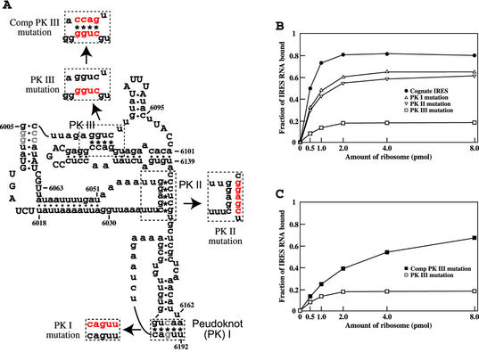
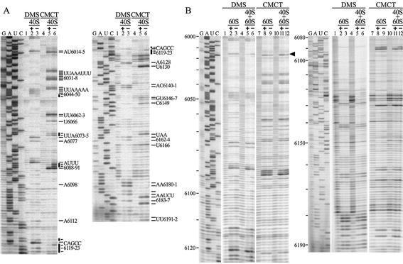
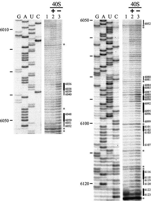
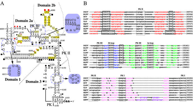
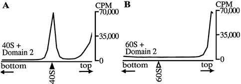
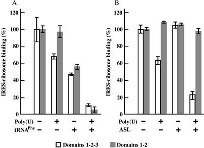
Plautia stali intestine virus (PSIV) has an internal ribosome entry site (IRES) at the intergenic region of the genome. The PSIV IRES initiates translation with glutamine rather than the universal methionine. To analyze the mechanism of IRES-mediated initiation, binding of IRES RNA to salt-washed ribosomes in the absence of translation factors was studied. Among the three pseudoknots (PKs I, II and III) within the IRES, PK III was the most important for ribosome binding. Chemical footprint analyses showed that the loop parts of the two stem-loop structures in Domain 2, which are highly conserved in related viruses, are protected by 40S but not by 60S ribosomes. Because PK III is close to the two loops, these structural elements were considered to be important for binding of the 40S subunit. Competitive binding analyses showed that the IRES RNA does not bind poly(U)-programmed ribosomes preincubated with tRNA(Phe) or its anticodon stem- loop (ASL) fragment. However, Domain 3-deleted IRES bound to programmed ribosomes preincubated with the ASL, suggesting that Domains 1 and 2 have roles in IRES binding to 40S subunits and that Domain 3 is located at the ribosome decoding site.
Figures
Similar articles
-
A tertiary structure model of the internal ribosome entry site (IRES) for methionine-independent initiation of translation.RNA. 2001 Feb;7(2):266-74. doi: 10.1017/s1355838201001741. RNA. 2001. PMID: 11233983 Free PMC article.
-
Binding mode of the first aminoacyl-tRNA in translation initiation mediated by Plautia stali intestine virus internal ribosome entry site.J Biol Chem. 2007 Mar 16;282(11):7770-6. doi: 10.1074/jbc.M610887200. Epub 2007 Jan 5. J Biol Chem. 2007. PMID: 17209036
-
Conditional rather than absolute requirements of the capsid coding sequence for initiation of methionine-independent translation in Plautia stali intestine virus.J Virol. 2003 Nov;77(22):12002-10. doi: 10.1128/jvi.77.22.12002-12010.2003. J Virol. 2003. PMID: 14581537 Free PMC article.
-
Divergent IRES elements in invertebrates.Virus Res. 2006 Jul;119(1):16-28. doi: 10.1016/j.virusres.2005.10.011. Epub 2005 Nov 22. Virus Res. 2006. PMID: 16307820 Review.
-
IRES-induced conformational changes in the ribosome and the mechanism of translation initiation by internal ribosomal entry.Biochim Biophys Acta. 2009 Sep-Oct;1789(9-10):558-70. doi: 10.1016/j.bbagrm.2009.06.001. Epub 2009 Jun 17. Biochim Biophys Acta. 2009. PMID: 19539793 Free PMC article. Review.
Cited by
-
Host and viral translational mechanisms during cricket paralysis virus infection.J Virol. 2010 Jan;84(2):1124-38. doi: 10.1128/JVI.02006-09. Epub 2009 Nov 4. J Virol. 2010. PMID: 19889774 Free PMC article.
-
Dynamics of IRES-mediated translation.Philos Trans R Soc Lond B Biol Sci. 2017 Mar 19;372(1716):20160177. doi: 10.1098/rstb.2016.0177. Philos Trans R Soc Lond B Biol Sci. 2017. PMID: 28138065 Free PMC article. Review.
-
Conservation and diversity among the three-dimensional folds of the Dicistroviridae intergenic region IRESes.J Mol Biol. 2007 Jul 27;370(5):856-69. doi: 10.1016/j.jmb.2007.04.076. Epub 2007 May 8. J Mol Biol. 2007. PMID: 17544444 Free PMC article.
-
Viral RNA pseudoknots: versatile motifs in gene expression and replication.Nat Rev Microbiol. 2007 Aug;5(8):598-610. doi: 10.1038/nrmicro1704. Nat Rev Microbiol. 2007. PMID: 17632571 Free PMC article. Review.
-
Viral IRES RNA structures and ribosome interactions.Trends Biochem Sci. 2008 Jun;33(6):274-83. doi: 10.1016/j.tibs.2008.04.007. Epub 2008 May 28. Trends Biochem Sci. 2008. PMID: 18468443 Free PMC article. Review.
References
Publication types
MeSH terms
Substances
LinkOut - more resources
Full Text Sources
Other Literature Sources
Miscellaneous
