. 2003 May 1;31(9):2451-9.
doi: 10.1093/nar/gkg342.

Stimulation of human DNA polymerase epsilon by MDM2

Affiliations

Stimulation of human DNA polymerase epsilon by MDM2

Hitomi Asahara et al. Nucleic Acids Res. .

Abstract

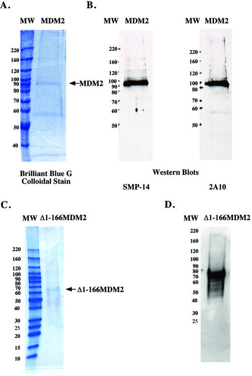
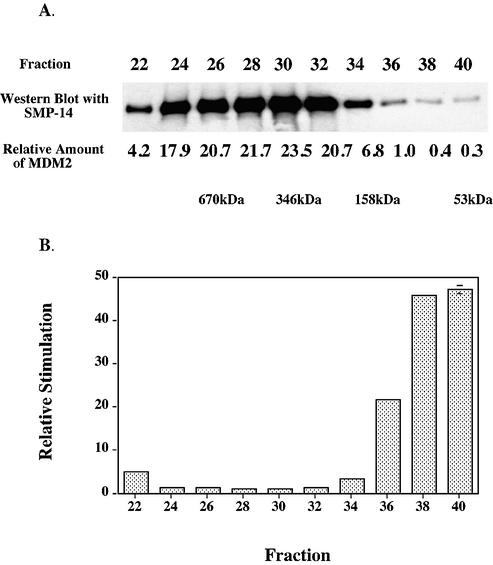
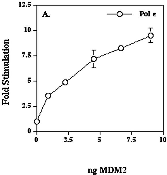
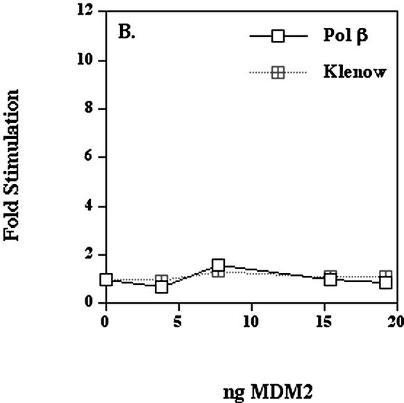
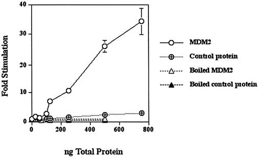
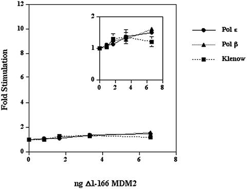
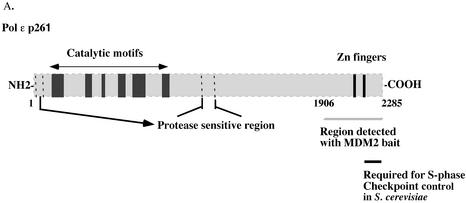
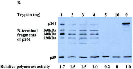
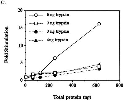
The human DNA polymerase epsilon catalytic subunit consists of a 140-kDa N-terminal domain that contains the catalytic activity and a 120-kDa C-terminal domain that binds to the other subunits and to exogenous peptides, including PCNA and MDM2. We report here that recombinant human MDM2 purified from insect cells or Escherichia coli stimulated the activity of DNA polymerase epsilon up to 10- and 40-fold, respectively, but not those of DNA polymerase beta or Klenow fragment of E.coli DNA polymerase I. Kinetic studies indicated that MDM2 increased the maximum velocity of the reaction, but did not change substrate affinities. The stimulation depended upon the interaction of the N-terminal 166 amino acid residues of MDM2 with the C-terminal domain of the full-length catalytic subunit, since the deletion of 166 amino acids from N-terminal of MDM2 or the removal of the C-terminal domain of DNA polymerase epsilon by trypsin digestion or competition for binding to it by the addition of excess C-terminal fragment eliminated the stimulation. Since DNA polymerase epsilon appears to be involved in DNA replication, recombination and repair synthesis, we suggest that MDM2 binding to DNA polymerase epsilon might be part of a reconfiguration process that allows DNA polymerase epsilon to associate with repair/recombination proteins in response to DNA damage.


Figures

Figure 1
Figure 1
SDS–PAGE of human MDM2 and Δ1–166 MDM2 expressed in insect cells. Purified MDM2 (roughly 0.6 µg of total protein) was subjected to SDS–PAGE on 7.5% polyacrylamide gels and then stained with Brilliant Blue G Colloidal stain (A) or electroblotted onto a nitrocellulose filter and probed with anti-MDM2 antibodies SMP-14 or 2A10 whose epitopes are amino acid 154–167 and 295–330, respectively (B). Purified Δ1–166 MDM2 was subjected SDS–PAGE on 4–20% linear gradient polyacrylamide gel and stained with Brilliant Blue G Colloidal stain (roughly 0.96 µg of total protein) (C) or electroblotted onto a nitrocellulose filter and probed with antibody 2A10 (roughly 0.48 µg of total protein) (D). Molecular weight markers were the Invitrogen BenchMark™ Protein Ladder.
Figure 2
Figure 2
Chromatography of recombinant MDM2 upon Superdex. MDM2 expressed in insect cells was purified through chromatography upon DEAE–Sephacel as described in Materials and Methods, and fractions containing MDM2 were concentrated using a Millipore Centriprep YM-50 and then chromatographed upon Superdex 200 HR 10/30 (Amersham Biosciences). (A) Equal volumes of samples were analyzed by immunoblots using antibody SMP-14. (B) The Superdex fractions were assayed for DNA polymerase ε simulating activity such that each fraction stimulated between 2- and 4-fold. The amounts of MDM2 in each fraction were quantitated from the immunoblot (with a lesser exposure time than the one shown) using the program ImageQuant for Macintosh v. 1.2. The fold-stimulations were then normalized for the amount of MDM2 antigen in each fraction and then those ratios were normalized to that of fraction 30 which had the highest amount of MDM2 antigen. The values are averages of duplicate assays and error bars were too small to show except for fraction 40. The Bio-Rad gel filtration standard (Catalog number 151-1901) was used to calibrate the column.
Figure 3
Figure 3
Human MDM2 expressed in insect cells stimulates human DNA polymerase ε. (A) DNA polymerase ε (5.8 × 10⁻³ units) was pre-incubated as indicated with the preparation of MDM2 shown in Figure 1A for 1 h on ice and then the activity was assayed as described in Materials and Methods. (B) The MDM2 was pre-incubated for 1 h on ice with either recombinant human DNA polymerase β expressed in E.coli (7.7 × 10⁻³ units) or Klenow fragment of E.coli DNA polymerase I (8.8 × 10⁻³ units) and then assayed. This preparation of MDM2 stimulated the activity of DNA polymerase ε up to 11-fold in the range of 4–20 ng. Values represent the mean of duplicate assays and in most cases error bars would be smaller than the data symbols.
Figure 3
Figure 3
Human MDM2 expressed in insect cells stimulates human DNA polymerase ε. (A) DNA polymerase ε (5.8 × 10⁻³ units) was pre-incubated as indicated with the preparation of MDM2 shown in Figure 1A for 1 h on ice and then the activity was assayed as described in Materials and Methods. (B) The MDM2 was pre-incubated for 1 h on ice with either recombinant human DNA polymerase β expressed in E.coli (7.7 × 10⁻³ units) or Klenow fragment of E.coli DNA polymerase I (8.8 × 10⁻³ units) and then assayed. This preparation of MDM2 stimulated the activity of DNA polymerase ε up to 11-fold in the range of 4–20 ng. Values represent the mean of duplicate assays and in most cases error bars would be smaller than the data symbols.
Figure 4
Figure 4
Human MDM2 expressed in E.coli stimulates HeLa pol ε. Pol ε (4.4–6 × 10⁻³ U was pre-incubated with the indicated amounts of MDM2, control protein or boiled MDM2 for 30 min on ice and then assayed as described in Materials and Methods. The MDM2 protein was about 5–10% of the total protein in the preparation. Values represent the mean of duplicate assays. In most cases, the error bars were smaller than the data symbols.
Figure 5
Figure 5
Effect of Δ1–166 MDM2 upon polymerase activities. DNA polymerase ε (12 × 10–3 U), DNA polymerase β (32 × 10–3 U) or Klenow fragment of E.coli DNA polymerase I (19 × 10–3 units) were pre-incubated with the indicated amounts of purified Δ1–166 MDM2 on ice for 1 h and then assayed. The inset is a magnification of the data. Values represent the mean of duplicate assays for DNA polymerase β and Klenow fragment and the mean of triplicate assays for DNA polymerase ε. Error bars were generally smaller than the data symbols.
Figure 6
Figure 6
The C-terminal domain of intact DNA polymerase ε p261 is essential for stimulation by MDM2. (A) HeLa p261 can be separated into two domains which are linked by a protease-sensitive region. The N‐terminal domain contains the catalytic motifs, whereas the C-terminal domain binds other subunits and proteins. (B and C) Fifty nanogram aliquots of purified HeLa DNA polymerase ε were digested with various amounts of trypsin as indicated for 15 min at 37°C, after which 1 µg of soybean trypsin inhibitor was added to stop each reaction. Half of each product was separated by 7.5% SDS–PAGE and then transferred to a nitrocellulose filter which was probed with anti-p261 antibodies 3C5.1, which recognizes the N‐terminal domain of p261, and 3A5.6, which recognizes p59. The remainder of each sample was assayed for polymerase activity in the absence (B) or presence (C) of recombinant MDM2 expressed in E.coli.
Figure 6
Figure 6
The C-terminal domain of intact DNA polymerase ε p261 is essential for stimulation by MDM2. (A) HeLa p261 can be separated into two domains which are linked by a protease-sensitive region. The N‐terminal domain contains the catalytic motifs, whereas the C-terminal domain binds other subunits and proteins. (B and C) Fifty nanogram aliquots of purified HeLa DNA polymerase ε were digested with various amounts of trypsin as indicated for 15 min at 37°C, after which 1 µg of soybean trypsin inhibitor was added to stop each reaction. Half of each product was separated by 7.5% SDS–PAGE and then transferred to a nitrocellulose filter which was probed with anti-p261 antibodies 3C5.1, which recognizes the N‐terminal domain of p261, and 3A5.6, which recognizes p59. The remainder of each sample was assayed for polymerase activity in the absence (B) or presence (C) of recombinant MDM2 expressed in E.coli.

Similar articles

Cited by

References

    1. Fakharzadeh S.S., Trusko,S.P. and George,D.L. (1991) Tumorigenic potential associated with enhanced expression of a gene that is amplified in a mouse tumor cell line. EMBO J., 10, 1565–1569. - PMC - PubMed
    1. Finlay C.A. (1993) The mdm-2 oncogene can overcome wild-type p53 suppression of transformed cell growth. Mol. Cell. Biol., 13, 301–306. - PMC - PubMed
    1. Oliner J.D., Kinzler,K.W., Meltzer,P.S., George,D.L. and Vogelstein,B. (1992) Amplification of a gene encoding a p53-associated protein in human sarcomas. Nature, 358, 80–83. - PubMed
    1. Maya R., Balass,M., Kim,S.T., Shkedy,D., Leal,J.F., Shifman,O., Moas,M., Buschmann,T., Ronai,Z., Shiloh,Y., Kastan,M.B., Katzir,E. and Oren,M. (2001) ATM-dependent phosphorylation of Mdm2 on serine 395: role in p53 activation by DNA damage. Genes Dev., 15, 1067–1077. - PMC - PubMed
    1. Haupt Y., Maya,R., Kazaz,A. and Oren,M. (1997) Mdm2 promotes the rapid degradation of p53. Nature, 387, 296–299. - PubMed

Publication types

MeSH terms