. 2003 Mar;202(Pt 3):269-77.
doi: 10.1046/j.1469-7580.2003.00158.x.

Expression of bone morphogenetic proteins and cartilage-derived morphogenetic proteins during osteophyte formation in humans

Affiliations

Expression of bone morphogenetic proteins and cartilage-derived morphogenetic proteins during osteophyte formation in humans

Sanja Zoricic et al. J Anat. 2003 Mar.

Abstract

Bone- and cartilage-derived morphogenetic proteins (BMPs and CDMPs), which are TGFbeta superfamily members, are growth and differentiation factors that have been recently isolated, cloned and biologically characterized. They are important regulators of key events in the processes of bone formation during embryogenesis, postnatal growth, remodelling and regeneration of the skeleton. In the present study, we used immunohistochemical methods to investigate the distribution of BMP-2, -3, -5, -6, -7 and CDMP-1, -2, -3 in human osteophytes (abnormal bony outgrowths) isolated from osteoarthritic hip and knee joints from patients undergoing total joint replacement surgery. All osteophytes consisted of three different areas of active bone formation: (1) endochondral bone formation within cartilage residues; (2) intramembranous bone formation within the fibrous tissue cover and (3) bone formation within bone marrow spaces. The immunohistochemistry of certain BMPs and CDMPs in each of these three different bone formation sites was determined. The results indicate that each BMP has a distinct pattern of distribution. Immunoreactivity for BMP-2 was observed in fibrous tissue matrix as well as in osteoblasts; BMP-3 was mainly present in osteoblasts; BMP-6 was restricted to young osteocytes and bone matrix; BMP-7 was observed in hypertrophic chondrocytes, osteoblasts and young osteocytes of both endochondral and intramembranous bone formation sites. CDMP-1, -2 and -3 were strongly expressed in all cartilage cells. Surprisingly, BMP-3 and -6 were found in osteoclasts at the sites of bone resorption. Since a similar distribution pattern of bone morphogenetic proteins was observed during embryonal bone development, it is suggested that osteophyte formation is regulated by the same molecular mechanism as normal bone during embryogenesis.


Figures

Fig. 1
Fig. 1
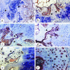
(a) Cross-section of typical osteophyte from hip joint, showing three main types of tissue and typical arrangement of the zones as distinguished by histomorphological criteria and olivine blue staining pattern: fibrous tissue (f), cartilage (c), bone (wb). The peripheral rim of the osteophyte merging with the synovial tissue (arrow) is covered by fibrous tissue and cross-passing blood vessels, with this fibrous tissue also extending over the surface layer of the cartilaginous tissue (toluidine blue, magnification 40×). (b) On the eroded side (arrow) of the cartilage (c), numerous osteoclasts, osteoblasts and highly cellular marrow stromal tissues were present; the newly formed woven bone trabeculae (wb) still contain areas of residual cartilaginous core (cc), clearly indicating endochondral bone development (toluidine blue, magnification 200×). (c) Intramembranous bone formation on the surface of the osteophyte; this fragment shows clusters of mesenchymal cells within connective fibrous (f) tissue that have gradually become enlarged and embedded in unmineralized woven bone matrix (wb), laying down the pre-existing mineralized lamellar bone trabeculae (lb) (toluidine blue, magnification 200×). (d) Specific area of active remodelling within osteophytes showed that within marrow containing connective tissue, there is differentiation of marrow cells into osteoblasts (arrow). As a result of this cell differentiation, new bone trabeculae arise on the free surface of old bone trabeculae. These two ‘types’ of trabeculaes can be distinguished, as shown clearly on this fragment found within the osteophyte (toluidine blue, magnification 200×).
Fig. 2
Fig. 2
BMPs immunostaining in the areas of endochondral bone development within osteophytes. (a) BMP-7 immunostaining was most intense in the cytoplasm of hypertrophic chondrocytes (arrowheads; magnification 100×). (b) Immunostaining in osteoblasts (arrowheads) of the newly formed woven bone trabeculae on the eroded side of the cartilage (magnification 200×). (c) BMP-3 immunostaining was found in some hypertrophic chondrocytes (white arrowheads), osteoblasts and young osteocytes (black arrowheads) (magnification 100×). (d) BMP-5 immunostaining was found in chondrocytes (white arrowheads) at the site of vascular invasion and matrix resorption, and in osteoblasts (black arrowheads) at the ossification front (magnification 100×). (e) BMP-6 immunostaining in osteoblasts (black arrowheads) and osteocytes (white arrowheads) of the newly formed woven bone trabeculae and in a minority of the chondrocytes (magnification 400×). (f) CDMP-1 immunostaining in osteophytes showed that all cartilaginous cells (black arrowheads) of the cartilage subjected to the endochondral bone development cascade were CDMP-1 positive; the covering fibrous tissue and underlying bone tissue showed no CDMP-1 immunostaining (magnification 400×).
Fig. 3
Fig. 3
(a) Matrix staining for BMP-2 was observed in the cells (arrowheads) of the superficial part of the covering fibrous tissue (asterisk) and in the matrix of the newly developed intramembranous bone (magnification 40×). (b) BMP-3-positive immunostaining in osteoblasts (arrowheads) at the sites of intramembranous bone formation (magnification 100×). BMP-3 positive (c, magnification 200×) and BMP-6 positive (d, magnification 400×) immunostaining were observed in osteoclasts (arrowheads) active in bone remodelling sites.

References

    1. Aigner T, Dietz U, Stöβ H, von der Mark K. Differential expression of collagen types I, II, III, and X in human osteophytes. Lab. Invest. 1995;73:236–243. - PubMed
    1. Anderson HC, Hodges PT, Moylan PE. Bone morphogenetic protein expression by cells of human and rat growth plate, metaphysis and articular cartilage. J. Bone Mineral Res. 1999;14(Suppl. 1):5309.
    1. Anderson HC, Hodges PT, Aguilera XM, Missana L, Moylan PE. Bone morphogenetic protein (BMP) localization in developing human and rat growth plate, metaphysis, epiphysis and articular cartilage. J. Histochem. Cytochem. 2000;48:1493–1502. - PubMed
    1. Aspenberg P, Basic N, Tagil M, Vukicevic S. Reduced expression of BMP-3 due to mechanical loading – A link between mechanical stimuli and tissue differentiation. Acta Orthopaedica Scand. 2000;71:558–562. - PubMed
    1. Bostrom MPG, Lane JM, Berberian WS, Missri AA, Tomin E, Weiland A, et al. Immunolocalization and expression of bone morphogenetic proteins-2 and -4 in fracture healing. J. Orthopaedic Res. 1995;13:357–367. - PubMed

MeSH terms