

Recurrent de novo point mutations in lamin A cause Hutchinson-Gilford progeria syndrome
- PMID: 12714972
- PMCID: PMC10540076
- DOI: 10.1038/nature01629
Recurrent de novo point mutations in lamin A cause Hutchinson-Gilford progeria syndrome
Abstract
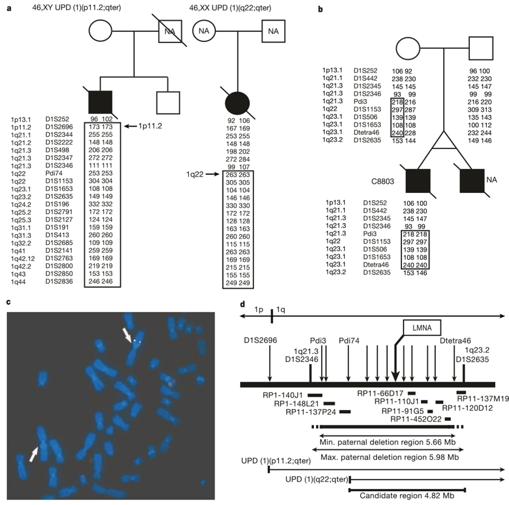
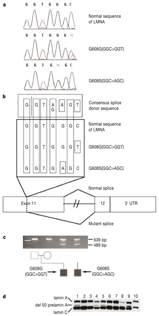
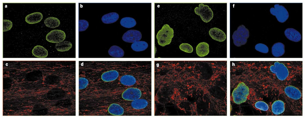
Hutchinson-Gilford progeria syndrome (HGPS) is a rare genetic disorder characterized by features reminiscent of marked premature ageing. Here, we present evidence of mutations in lamin A (LMNA) as the cause of this disorder. The HGPS gene was initially localized to chromosome 1q by observing two cases of uniparental isodisomy of 1q-the inheritance of both copies of this material from one parent-and one case with a 6-megabase paternal interstitial deletion. Sequencing of LMNA, located in this interval and previously implicated in several other heritable disorders, revealed that 18 out of 20 classical cases of HGPS harboured an identical de novo (that is, newly arisen and not inherited) single-base substitution, G608G(GGC > GGT), within exon 11. One additional case was identified with a different substitution within the same codon. Both of these mutations result in activation of a cryptic splice site within exon 11, resulting in production of a protein product that deletes 50 amino acids near the carboxy terminus. Immunofluorescence of HGPS fibroblasts with antibodies directed against lamin A revealed that many cells show visible abnormalities of the nuclear membrane. The discovery of the molecular basis of this disease may shed light on the general phenomenon of human ageing.
Conflict of interest statement
Figures
References
-
- DeBusk FL The Hutchinson-Gilford progeria syndrome. J. Pediat 80, 697–724 (1972). - PubMed
-
- Baker PB, Baba N & Boesel CP Cardiovascular abnormalities in progeria. Case report and review of the literature. Arch. Pathol. Lab. Med 105, 384–386 (1981). - PubMed
-
- Burke B & Stewart CL Life at the edge: the nuclear envelope and human disease. Nature Rev. 3, 575–585 (2002). - PubMed
-
- Genschel J & Schmidt H-J Mutations in the LMNA gene encoding lamin A/C. Hum. Mutat 16, 451–459 (2000). - PubMed
-
- Brown WT Human mutations affecting aging—a review. Mech. Aging Dev 9, 325–336 (1979). - PubMed
Publication types
MeSH terms
Substances
Grants and funding
LinkOut - more resources
Full Text Sources
Other Literature Sources
Medical
Molecular Biology Databases
Research Materials
Miscellaneous
