. 2003 Apr 15;23(8):3234-42.
doi: 10.1523/JNEUROSCI.23-08-03234.2003.

Cell type-specific roles for tissue plasminogen activator released by neurons or microglia after excitotoxic injury

Affiliations

Cell type-specific roles for tissue plasminogen activator released by neurons or microglia after excitotoxic injury

Chia-Jen Siao et al. J Neurosci. .

Abstract

Tissue plasminogen activator (tPA) plays important roles in the brain after excitotoxic injury. It is released by both neurons and microglia and mediates neuronal death and microglial activation. Mice lacking tPA are resistant to excitotoxicity and show very limited microglial activation. Activated microglia are neurotoxic in culture, but this phenomenon is not well documented in vivo. To further understand the sequence of events through which tPA mediates microglial activation and neurodegeneration, we have generated mice that exhibit restricted expression of tPA through introduction of tPA transgenes under the control of neuronal- or microglial-specific promoters into tPA-deficient mice. Neither strain of transgenic mice shows abnormal brain morphology or inflammation in the absence of injury, and unilateral intrahippocampal kainate injections into the transgenic mice induced excitotoxicity and microglial activation reminiscent of wild-type mice. However, there are differences in the kinetics of the resulting pathology. The neuronal tPA-expressing mice exhibit accelerated microglial activation compared with wild-type or microglial tPA-expressing mice. However, microglial tPA-expressing mice exhibit greater neurodegeneration. These data suggest a model in which tPA plays different roles after kainate injection depending on whether it is released by neurons or microglia. We propose that tPA, initially secreted from injured neurons, acts as a cytokine to activate microglia at the site of injury. These activated microglia then secrete additional tPA, which promotes extracellular matrix degradation, neurodegeneration, and self-proliferation. We suggest that an approach to attenuate microglia-mediated neuronal death in vivo might be to pharmacologically prevent microglial activation.


Figures

Fig. 1.
Fig. 1.
Characterization of transgenic mice.a, A neuron-specific transgenic construct. The promoter for the neurofilament light chain gene was fused to mtPA, and relevant restriction enzyme sites are labeled. b, A microglia-specific transgenic construct. The promoter for the gene encoding the receptor for M-CSF (also known as c-fms) was fused to mouse tPA cDNA. c, Genotyping PCR results for NF-L-tPA founder mice. −, No DNA; p, transgenic plasmid; tg−, nontransgenic littermate control. The primers that amplify the transgene (5′ primer in the promoter and 3′ primer in tPA cDNA) specifically produce a 512 bp product, whereas the primers for tPA cDNA amplify a 470 bp product.d, Genotyping PCR results for Fms-tPA founder mice. The transgene-specific promoters amplified a 590 bp product.e, The transgenic mice express cell-specific tPA. Primary microglia or neurons were cultured as described, and then total RNA was extracted and reverse transcribed. Primers for BDNF (B, neuronal), F4/80 (F, microglial), or tPA (t) were used to amplify specific products to show that the cells express tPA only from neurons (NF-L-tPA mice) or microglia (Fms-tPA mice). f,In situ hybridization results for transgenic mice. The NF-L-tPA section shows a strong tPA RNA presence in the CA1 pyramidal neurons. The Fms-tPA sections show a more diffuse tPA RNA presence around the CA3 subfield (arrowheads) as well as in the CC (arrowheads). Original magnification, 100×.
Fig. 2.
Fig. 2.
tPA secreted by transgenic mice is active. Anin situ zymographic assay was used on sections from wild-type (a), tPA−/−(b), NF-L-tPA (c), and Fms-tPA (d) mice to visualize endogenous tPA proteolytic activity. All sections except for tPA−/− showed tPA activity in the CA3/hilus regions of the hippocampus (arrows). The arrows in dalso indicate the stratum radiatum. Endogenous uPA activity was inhibited with amiloride. Original magnification, 10×.
Fig. 3.
Fig. 3.
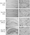
Transgenic mice show no neuronal death or microglial activation without injury. Cresyl violet stained hippocampal sections show an intact CA pyramidal neuronal layer in mice that were untreated (a, c) or injected with PBS only (e, g). b,d, f, h, In addition, sections (boxed insets) from the same mice were immunodetected with F4/80 antibody, which recognizes microglia/macrophages; the lack of specific staining shows that the brain parenchyma is not injured. Dorsal is at the top of all panels. Original magnifications: cresyl violet panels, 20×; immunohistochemistry panels, 400×. The high-magnification panels are from the CA1 subfield of the hippocampus. uninj., Uninjected; inj., injected.
Fig. 4.
Fig. 4.
Characterization of neuronal death in wild-type and transgenic mice after excitotoxicity. Mice were injected with KA and then killed at the indicated time points. Neuronal death was determined using cresyl violet, which outlines an intact pyramidal neuronal layer. Lack of staining indicates that the cells are dead.A, Left column, wild-type (wt) sections; middle column, NF-L-tPA sections; right column, Fms-tPA sections. Dorsal is at the top of all panels. Insets, Higher magnification of the CA1 subfield showing shrunken soma of dead neurons compared with larger stronger-staining cell bodies of neurons that survived the KA injection. Original magnification: large panels, 10×; insets, 200×. The hippocampal measurements were performed by investigators blind to the genotypes of the mice as well as to the time points after KA injection for each sample. B, A response comparable with that seen for KA injection is observed in the other transgenic lines as well at all time points. The sections presented are from 1 d after KA injection.
Fig. 5.
Fig. 5.
Quantification of neuronal death after KA injection. Sections treated as in Figure 4 were grouped by time points. The amount of neurodegeneration was measured using Scion Image and presented as the percentage of total hippocampal length of the ipsilateral injected side (SEM). These data are graphically presented as well. Note that the NF-L-tPA mice show a slower increase of neuronal loss at early time points (up to 2 d) compared with wild-type or Fms-tPA mice, although they match the percentage lost by Fms-tPA mice by 5 d.
Fig. 6.
Fig. 6.
Characterization of microglial activation in wild-type (wt) and transgenic mice after excitotoxicity. Mice were processed as described in Materials and Methods and Figure 4. Sections were immunodetected using either 5D4 (6 hr to 1 d) or F4/80 (2–5 d) antibodies. Note that even by 12 hr some activated microglia are visible, although not in the proximity of dying neurons in the CA1 field. Both antibodies recognize antigen in the microvasculature (small arrows), but activated microglia are clearly defined (large arrows, insets). Left column, Wild-type sections; middle column, NF-L-tPA sections; right column, Fms-tPA sections. Dorsal is located at the top of each panel. Original magnification: large panels, 200×; insets, 400×.

References

    1. Akassoglou K, Probert L, Kontogeorgos G, Kollias G. Astrocyte-specific but not neuron-specific transmembrane TNF triggers inflammation and degeneration in the central nervous system of transgenic mice. J Immunol. 1997;158:438–445. - PubMed
    1. Akiyama H, Nishimura T, Kondo H, Ikeda K, Hayashi Y, McGeer P. Expression of the receptor for macrophage colony stimulating factor by brain microglia and its upregulation in brains of patients with Alzheimer's disease and amyotrophic lateral sclerosis. Brain Res. 1994;639:171–174. - PubMed
    1. Andersson P, Perry V, Gordon S. The kinetics and morphological characteristics of the macrophage-microglial response to kainic acid-induced neuronal degeneration. Neuroscience. 1991;42:201–214. - PubMed
    1. Banati R, Graeber M. Surveillance, intervention and cytotoxicity: is there a protective role of microglia? Dev Neurosci. 1994;16:114–127. - PubMed
    1. Baranes D, Lederfein D, Huang YY, Chen M, Bailey C, Kandel E. Tissue plasminogen activator contributes to the late phase of LTP and to synaptic growth in the hippocampal mossy fiber pathway. Neuron. 1998;21:813–825. - PubMed

Publication types

MeSH terms