. 2003 Apr 15;23(8):3394-406.
doi: 10.1523/JNEUROSCI.23-08-03394.2003.

Coordinate regulation of glutathione biosynthesis and release by Nrf2-expressing glia potently protects neurons from oxidative stress

Affiliations

Coordinate regulation of glutathione biosynthesis and release by Nrf2-expressing glia potently protects neurons from oxidative stress

Andy Y Shih et al. J Neurosci. .

Abstract

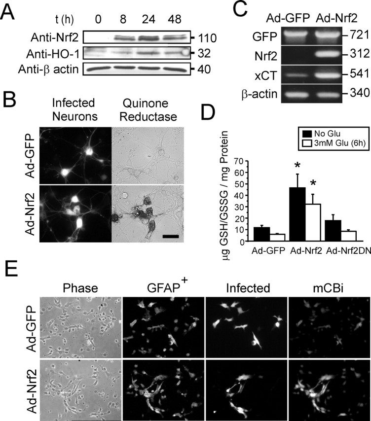
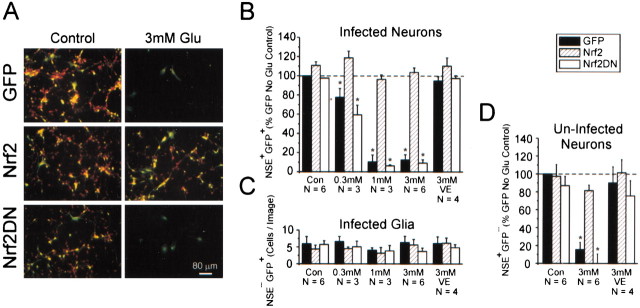
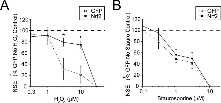
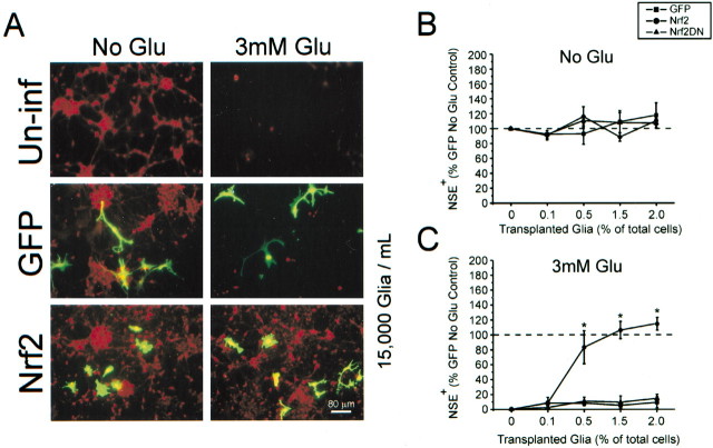
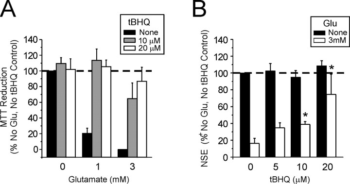
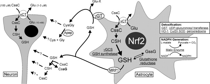
Astrocytes have a higher antioxidant potential in comparison to neurons. Pathways associated with this selective advantage include the transcriptional regulation of antioxidant enzymes via the action of the Cap'n'Collar transcription factor Nrf2 at the antioxidant response element (ARE). Here we show that Nrf2 overexpression can reengineer neurons to express this glial pathway and enhance antioxidant gene expression. However, Nrf2-mediated protection from oxidative stress is conferred primarily by glia in mixed cultures. The antioxidant properties of Nrf2-overexpressing glia are more pronounced than those of neurons, and a relatively small number of these glia (< 1% of total cell number added) could protect fully cocultured naive neurons from oxidative glutamate toxicity associated with glutathione (GSH) depletion. Microarray and biochemical analyses indicate a coordinated upregulation of enzymes involved in GSH biosynthesis (xCT cystine antiporter, gamma-glutamylcysteine synthetase, and GSH synthase), use (glutathione S-transferase and glutathione reductase), and export (multidrug resistance protein 1) with Nrf2 overexpression, leading to an increase in both media and intracellular GSH. Selective inhibition of glial GSH synthesis and the supplementation of media GSH indicated that an Nrf2-dependent increase in glial GSH synthesis was both necessary and sufficient for the protection of neurons, respectively. Neuroprotection was not limited to overexpression of Nrf2, because activation of endogenous glial Nrf2 by the small molecule ARE inducer, tert-butylhydroquinone, also protected against oxidative glutamate toxicity.


Figures

Fig. 1.
Fig. 1.
Schematic explanation of neuron–glial coculture setups used in this study. A, For a simple neuron–glial coculture setup the virus-infected glia were transplanted directly into naive (no contact with virus) mixed neuronal–glial cultures after 24 hr of transgene expression. B, Some experiments required a setup by which glia were separated physically from neurons (membrane-delimited coculture). This system consisted of naive cultures prepared in 24-well plates; infected glia were separated by a culture plate insert. Both cocultures were maintained for 24 hr and then exposed to 3 mm glutamate (Glu) for a further 24 hr, followed by quantitation of neuronal viability.
Fig. 2.
Fig. 2.
Cortical glia have higher basal Nrf2 expression and ARE promotor activity than neurons. A, Western blot of heterologously expressed Nrf2 (105 kDa) and Nrf2DN (28 kDa) in HEK293 cells. B, A comigrating 105 kDa band corresponding to Nrf2 is observed in enriched cortical glial, but not neuronal, cultures. Densitometric analysis reveals an ∼12-fold difference in Nrf2 protein (n = 3); *p < 0.05. C, Coexpression of Nrf2 cDNA with a hPAP-encoding reporter of ARE-mediated gene expression (rQR51) greatly increases neuronal hPAP expression. Reporter constructs carrying a mutation within the core ARE consensus sequence (rQR51Mut) were not inducible. Coexpression with Nrf2DN cDNA suppresses both neuronal and glial hPAP expression. *p < 0.05, neuron comparison to pEF (empty vector) control; #p< 0.05, glial comparison to pEF control. D, E,Representative hPAP-stained astrocyte-like cells with coexpression of pEF vector only. F, G, With Nrf2 overexpression cells of both neuronal and glial morphology show ARE-driven hPAP expression. Data from hPAP experiments represent the mean ± SEM number of cells counted from triplicate coverslips over four independent experiments.
Fig. 3.
Fig. 3.
Ad-Nrf2-infected cultures exhibit enhanced antioxidant potential. A, Time course evaluation of Nrf2 protein overexpression with parallel induction of HO-1 expression.B, Histochemical staining revealed increased NQO1 activity in ad-Nrf2-infected neurons, but not neighboring uninfected neurons, visible under DIC optics (bottom panels). Neuronal NQO1 staining was not observed in the ad-GFP group (top panels). Scale bar, 20 μm. C, xCT mRNA levels increase with Nrf2 overexpression as shown by RT-PCR. Nrf2 mRNA derived from infection was detected by using selective mouse primers. D, Nrf2 overexpression increases total intracellular GSH/GSSG levels. Sublethal glutamate exposure (6 hr) leads to partial depletion of intracellular GSH in all groups (open bars). Control groups represent a separate group of cultures with no glutamate exposure but that were vehicle-treated (filled bars). Culture were given a total of 48 hr for expression before being harvested for GSH analysis. E,Increase in mCBi staining is primarily enriched in glia of ad-Nrf2-infected mixed cultures. Cultures are depicted in phase-contrast (Phase), fluorescence immunostaining for anti-GFAP (GFAP+) and anti-GFP (Infected), and 60 μm mCBi staining (mCBi). mCBi staining images show selected areas with high numbers of glial clusters and are not representative of actual mixed culture composition. HO-1 and NQO1 images are representative of at least three separate experiments. GSH data represent mean ± SEM of four separate experiments performed in duplicate; *p < 0.05, compared with GFP control.
Fig. 4.
Fig. 4.
Nrf2 overexpression in a subpopulation of cells confers widespread neuronal protection from oxidative glutamate toxicity. A, Immunocytochemistry for eGFP (green, identifying infected cells) and NSE (red marker, a neuron-selective marker). Within a typical ad-GFP-infected culture the infected neurons (yellow, red + green), uninfected neurons (red), and infected glia (green) can be observed. B, Group data evaluating the vulnerability of infected neurons to oxidative glutamate toxicity. Data are expressed as a percentage of GFP+NSE+ cells (presumed infected neurons) in the indicated glutamate treatment group as compared with the ad-GFP control group. VE, Vitamin E (α-tocopherol), 100 μm.C, Viability of GFP+NSE cells (presumed infected glia) present per image was not affected significantly with glutamate treatment.D, Uninfected neurons within cultures containing Nrf2-infected cells are more resistant to oxidative glutamate toxicity. Data represent the mean ± SEM number of cells counted over triplicate wells from at least three independent experiments; *p < 0.05, compared with the GFP control no-glutamate group.
Fig. 5.
Fig. 5.
Nrf2 overexpression in mixed immature cortical cultures protects neurons from H2O2-mediated toxicity, but not staurosporine-induced apoptosis. Ad-GFP- and ad-Nrf2-infected cultures were allowed to express transgenes for 48 hr before exposure to 0.3–30 μm H2O2 (A;n = 3) or 0.1–10 μm staurosporine (B; n = 4) for a further 24 hr. Cells were stained for NSE to evaluate neuronal viability. Data represent the mean ± SEM from the indicated number of experiments performed in quadruplicate; *p < 0.05, compared with ad-GFP-infected control.
Fig. 6.
Fig. 6.
A small fraction of infected glial cells is sufficient to protect neurons from oxidative glutamate toxicity.A, Representative images from glial–neuron coculture setup (see Fig. 1A). Within a typical coculture infected glia (green) and uninfected neurons (red) can be observed. Uninf, Uninfected glia transplanted. B, Group data obtained from plate scanning for NSE (red) fluorescence.C, Decreased neuronal viability is demonstrated by a loss of red fluorescence. The addition of Nrf2-overexpressing glia restores NSE expression to levels found in an ad-GFP-infected group that was not exposed to glutamate. Data represent the mean ± SEM from three separate experiments performed in quadruplicate; *p < 0.05. Scale bar, 80 μm.
Fig. 7.
Fig. 7.
Release of GSH from glia is both sufficient and necessary for conferring neuronal protection. A,B, Infection with ad-Nrf2 increases total intracellular GSH/GSSG as well as GSH released into the medium (MEM/5% FCS, no phenol red). C, Exogenous addition of reduced GSH concurrently with glutamate treatment protects neurons from oxidative glutamate toxicity. D, Glial GSH release is necessary for Nrf2-dependent neuronal protection. A membrane-delimited coculture (see Fig. 1B) was used, allowing enriched glial cultures to be pretreated separately with the GSH synthesis inhibitor BSO and then to be washed and added to wells containing neurons. BSO pretreatment (200 μm) for 24 hr produces long-term reduction of intracellular GSH/GSSG and release of GSH from glia and abolishes glial-mediated neuronal protection. Data represent the mean ± SEM of at least three independent experiments performed in triplicate; #*p < 0.05.
Fig. 8.
Fig. 8.
Neuronal protection can be achieved by activation of endogenous Nrf2 with the use of a small molecule inducer.A, Immature cortical cultures pretreated for 24 hr with 10 and 20 μm tBHQ are protected from 1–3 mmglutamate exposure. Partial protection is conferred by tBHQ treatment at 3 mm glutamate. B, Selective induction of endogenous Nrf2 in glia led to partial neuronal protection from oxidative glutamate toxicity. Glia pretreated with a range of tBHQ concentrations (0–20 μm) for 24 hr were transplanted into naive neuronal cultures at a plating density of 5% of the total cell number, using a coculture setup (see Fig.1A). Data represent the mean ± SEM from three independent experiments performed in quadruplicate; *p < 0.05.
Fig. 9.
Fig. 9.
Schematic diagram of GSH biosynthesis and release pathways that may be involved with Nrf2-dependent coupling of GSH between astrocytes and neurons. Astrocyte GSH synthesis and release are more robust with Nrf2 overexpression. Higher levels of xCT (system xc) expression were detected, which promotes cystine uptake and provides a precursor for GSH synthesis. Microarray analyses indicate that all major enzymes involved in GSH biosynthesis are upregulated also. GSH release from astrocytes is increased, leading into several possible pathways of extracellular GSH metabolism, including an initial breakdown by γ-glutamylcysteine transpeptidase (γGT) and possible further breakdown by aminopeptidase (Apep) into the glutathione precursors Cys, Gly, and CysGly suitable for neuronal uptake (Dringen et al., 1999, 2001). Alternatively, extracellular GSH may be taken up by neurons directly or may contribute to the reduction of cystine into cysteine, which may be a source of sulfhydryl species for neuronal uptake (Sagara et al., 1993;Wang and Cynader, 2000). Neurons may also uptake cystine for glutathione synthesis (Murphy et al., 1990). Microarray analyses also indicate the upregulation of additional factors involved in detoxification, ROS scavenging, and NADPH production that may work together with GSH to protect neurons. Genes that are upregulated significantly by Nrf2 overexpression are underlined. Intracellular and extracellular concentrations of glutamate are average values from a combination of previous studies in the human brain and CSF (Siegel, 1981). CSF levels of cystine are typically very low at ∼0.2 μm (Lakke and Teelken, 1976; Araki et al., 1988; Wang and Cynader, 2000). The Invitrogen MEM used in this study is formulated to contain 100 μm cystine.

References

    1. Ahlgren-Beckendorf JA, Reising AM, Schander MA, Herdler JW, Johnson JA. Coordinate regulation of NAD(P)H:quinone oxidoreductase and glutathione S-transferases in primary cultures of rat neurons and glia: role of the antioxidant/electrophile responsive element. Glia. 1999;25:131–142. - PubMed
    1. Alam J, Stewart D, Touchard C, Boinapally S, Choi AM, Cook JL. Nrf2, a Cap'n'Collar transcription factor, regulates induction of the heme oxygenase-1 gene. J Biol Chem. 1999;274:26071–26078. - PubMed
    1. Araki K, Harada M, Ueda Y, Takino T, Kuriyama K. Alteration of amino acid content of cerebrospinal fluid from patients with epilepsy. Acta Neurol Scand. 1988;78:473–479. - PubMed
    1. Bannai S. Exchange of cystine and glutamate across plasma membrane of human fibroblasts. J Biol Chem. 1986;261:2256–2263. - PubMed
    1. Bloom D, Dhakshinamoorthy S, Jaiswal AK. Site-directed mutagenesis of cysteine to serine in the DNA binding region of Nrf2 decreases its capacity to upregulate antioxidant response element-mediated expression and antioxidant induction of NAD(P)H:quinone oxidoreductase1 gene. Oncogene. 2002;21:2191–2200. - PubMed

Publication types

MeSH terms