. 2003 May 13;100(10):6109-14.
doi: 10.1073/pnas.1031395100. Epub 2003 Apr 25.

Heterogeneous nuclear ribonucleoprotein (hnRNP) binding to hormone response elements: a cause of vitamin D resistance

Affiliations

Heterogeneous nuclear ribonucleoprotein (hnRNP) binding to hormone response elements: a cause of vitamin D resistance

Hong Chen et al. Proc Natl Acad Sci U S A. .

Abstract

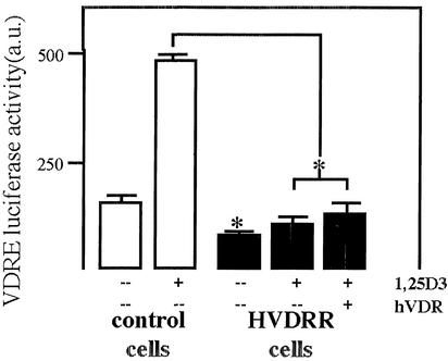
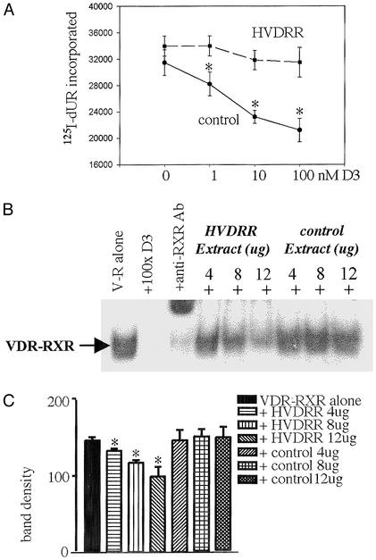
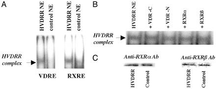
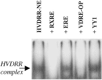
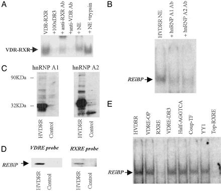
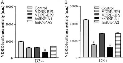
In previous studies, we have shown that steroid hormone resistance in New World primates occurs in the absence of abnormal expression of cognate nuclear receptors. Rather, these animals have elevated levels of heterogeneous nuclear ribonucleoproteins (hnRNPs) that act as hormone response element-binding proteins and attenuate target gene transactivation. Here we present evidence for a similar mechanism in humans via a patient with resistance to the active form of vitamin D [1,25-dihydroxyvitamin D(3) (1,25(OH)(2)D(3))] who presented with normal vitamin D receptor (VDR) expression. Initial cotransfection studies showed that the cells of the patient suppressed basal and hormone-induced transactivation by wild-type VDR. Electrophoretic mobility-shift assays and Western/Southwestern blot analyses indicated that this suppressive effect was due to overexpression of a nuclear protein that specifically interacts with a DNA response element known to bind retinoid X receptor-VDR heterodimers. Ab blocking in electrophoretic mobility-shift assays indicated that this dominant-negative acting protein was in the hnRNPA family of nucleic acid-binding proteins. Further studies have shown that several members of this family, most notably hnRNPA1, were able to suppress basal and 1,25(OH)(2)D(3)-induced luciferase activity. We therefore propose that this case of vitamin D resistance in a human subject is similar to that previously described for New World primates in which abnormal expression of a hormone response element-binding protein can cause target cell resistance to 1,25(OH)(2)D(3). That this protein is a member of the hnRNP family capable of interacting with double-stranded DNA highlights a potentially important new component of the complex machinery required for steroid hormone signal transduction.


Figures

Figure 1
Figure 1
VDR-mediated transactivation is suppressed in cells from a patient with HVDRR. Primary fibroblast cultures from a patient with HVDRR and an age- and sex-matched control were transfected with a VDRE-DR3 luciferase promoter–reporter construct in the presence or absence of 1,25(OH)2D3 (D3) (10 nM) and/or cotransfected wild-type VDR cDNA. Data are shown as arbitrary luciferase units (a.u.) relative to internal standard, β-galactosidase. Values are the mean ± SD of three separate assays. *, statistically different from control, P < 0.01.
Figure 2
Figure 2
Vitamin D resistance in HVDRR cells is associated with antagonism of normal VDR–RXR–VDRE nuclear complex formation. (A) Transformed B cells from the patient with HVDRR are resistant to 1,25(OH)2D3. Control and HVDRR lymphocytes were treated with the mitogen phorbol 12-myristate 13-acetate (PMA; 10 nM) in the presence or absence of increasing doses of 1,25(OH)2D3 (1–100 nM) for 72 h. Cell proliferation was then assessed by nuclear incorporation of [125I]iododeoxyuridine ([125I]dUR). *, statistically different from PMA-only controls cells in unpaired t tests, P < 0.01. (B) NEs from the HVDRR cells compete with VDR–RXR for association with a VDRE. Addition of increasing amounts of NEs from HVDRR cells (fourth through sixth lanes) but not the vitamin D-responsive control subject (seventh through ninth lanes) competed away recombinant human VDR–RXR binding to a labeled VDRE-DR3 double-stranded probe in EMSA (lane 1, V-R alone). Binding was also displaced by a 100-fold excess of unlabeled VDRE-DR3 (lane 2, +100 × DR3) and anti-RXR Ab (lane 3, +anti-RXR Ab). (C) Densitometric analysis of band density in replicate EMSAs (n = 3) representative assay shown in B (mean ± SD). Significant (P < 0.05) diminishment of probe-VDR–RXR complex formation with increasing doses of HVDRR NEs was achieved compared with equivalent control extracts.
Figure 3
Figure 3
NEs from HVDRR cells harbor a non-VDR, non-RXR-related REBiP. (A) EMSA analysis with consensus VDRE-DR3 and a consensus RXRE as probe incubated with NEs from HVDRR and control B cells. (B) Lack of EMSA supershift response by the RXRE-bound HVDRR NE complex in the presence of anti-VDR-C-terminal Ab (+VDR-C), anti-VDR-N-terminal Ab (+VDR-N), anti-RXRα Ab (+RXRα), and anti-RXRβ Ab (+RXRβ). (C) Western blot analyses showing normal expression of RXRα and RXRβ in NEs from control and HVDRR B cells.
Figure 4
Figure 4
EMSA analysis of RE sequence specificity for DNA complex formation by HVDRR extracts. NEs from HVDRR B cells (HVDRR-NE) were incubated with radiolabeled double-stranded RXRE probe in the absence (HVDRR-NE) or presence of an excess of unlabeled competitor probes. VDRE-OP, osteopontin VDRE; YY1, Ying Yang 1 transcription factor RE.
Figure 5
Figure 5
The REBiP present in HVDRR cells is a member of the hnRNP family. (A) The ability of HVDRR NE to compete out VDR–RXR complex formation with a VDRE-DR3 probe is abrogated by enzyme digestion with trypsin. EMSA lanes are as follows: 1, VDR–RXR alone; 2, VDR–RXR with 100-fold excess unlabeled probe; 3, VDR–RXR with anti-RXR Ab; 4, VDR–RXR with anti-VDR Ab; 5, VDR–RXR with untreated HVDRR NE; and 6, VDR–RXR with HVDRR NE pretreated with 0.5 μg/μl trypsin for 5 min followed by inactivation of trypsin with BSA 14 mg/ml. (B) HVDRR NE complex formation with VDRE-DR3 is blocked by anti-hnRNPA1 and anti-hnRNPA2 Abs. (C) Western blot analysis of hnRNPA1 (Left) and hnRNPA2 (Right) in HVDRR and control lymphocytes. (D) Southwestern blot analysis of HVDRR lymphocytes identifies a protein that associates with RXRE and VDRE-DR3. NEs from HVDRR and control cells were separated by PAGE and probed with radiolabeled RXRE or VDRE-DR3. (E) REBiP can bind to single- and double-stranded DNA. In competition EMSAs, HVDRR NEs bound to a single-strand labeled probe corresponding to the upper strand of the RXRE and was competed out by an excess of unlabeled double-stranded DNA probes.
Figure 6
Figure 6
hnRNPs antagonize VDRE-mediated transactivation. VDR-positive human kidney cells (HKC-8) were cotransfected with a VDRE-luciferase reporter plasmid together with expression constructs for hnRNPA1, hnRNPA2, VDRE-BP1 (NWP hnRNPA1), VDRE-BP2 (NWP hnRNPA2), or vector alone (control). Cells were cultured in the absence (A) or presence (B) of 10 nM 1,25(OH)2D3 for 24 h. Data are shown as arbitrary luciferase units (a.u.) relative to internal standard β-galactosidase measurements. Values are the mean ± SD of three separate assays. *, statistically different from equivalent control cells, P < 0.0001.

Similar articles

Cited by

References

    1. Chrousos G P, MacLusky N J, Brandon D D, Tomita M, Renquist D M, Loriaux D L, Lipsett M B. Adv Exp Med Biol. 1986;196:317–328. - PubMed
    1. Auchus R J, Fuqua S A. Semin Cell Biol. 1994;5:127–136. - PubMed
    1. Ing N H, O'Malley B W. In: Molecular Endocrinology: Basic Concepts and Clinical Correlations. Weintraub B D, editor. New York: Raven; 1995. pp. 195–215.
    1. Chen J D, Li H. Crit Rev Eukaryotic Gene Expression. 1998;8:169–190. - PubMed
    1. Freedman L P. Cell. 1999;97:5–8. - PubMed

Publication types