. 2003 May;12(5):893-902.
doi: 10.1110/ps.0237603.

Energy-dependent degradation: Linkage between ClpX-catalyzed nucleotide hydrolysis and protein-substrate processing

Affiliations

Energy-dependent degradation: Linkage between ClpX-catalyzed nucleotide hydrolysis and protein-substrate processing

Randall E Burton et al. Protein Sci. 2003 May.

Abstract

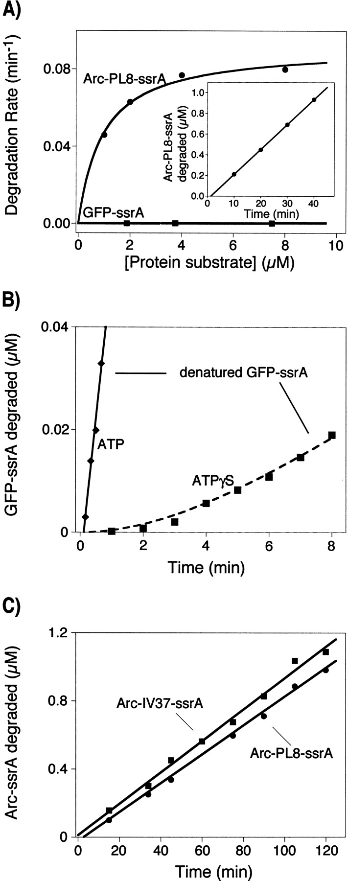
ClpX requires ATP to unfold protein substrates and translocate them into the proteolytic chamber of ClpP for degradation. The steady-state parameters for hydrolysis of ATP and ATPgammaS by ClpX were measured with different protein partners and the kinetics of degradation of ssrA-tagged substrates were determined with both nucleotides. ClpX hydrolyzed ATPgammaS to ADP and thiophosphate at a rate (6/min) significantly slower than ATP hydrolysis (140/min), but the hydrolysis of both nucleotides was increased by ssrA-tagged substrates and decreased by ClpP. K(M) and k(cat) for hydrolysis of ATP and ATPgammaS were linearly correlated over a 200-fold range, suggesting that protein partners largely affect k(cat) rather than nucleotide binding, indicating that most bound ATP leaves the enzyme by hydrolysis rather than dissociation, and placing an upper limit of approximately 15 micro M on K(D) for both nucleotides. Competition studies with ClpX and fluorescently labeled ADP gave inhibition constants for ATPgammaS ( approximately 2 micro M) and ADP ( approximately 3 micro M) under the reaction conditions used for steady-state kinetics. In the absence of Mg(2+), where hydrolysis does not occur, the inhibition constant for ATP ( approximately 55 micro M) was weaker but very similar to the value for ATPgammaS ( approximately 45 micro M). Compared with ATP, ATPgammaS supported slow but roughly comparable rates of ClpXP degradation for two Arc-ssrA substrates and denatured GFP-ssrA, but not of native GFP-ssrA. These results show that the processing of protein substrates by ClpX is closely coupled to the maximum rate of nucleotide hydrolysis.


Figures

Figure 1.
Figure 1.
(A) 31P-NMR spectra of 5 mM ATPγS (black) in lysis buffer with 10% D2O and 10 mM TMSP. The gray spectrum was recorded 21 h after addition of 0.3 μM ClpX6 to the sample. Resonances were assigned based on literature values (Eckstein and Goody 1976); (inset) Downfield resonance of the thiophosphate. (B) Time course of ATPγS hydrolysis. 31P-NMR peaks were assigned to the α (open circles) and β (open squares) phosphates of ATPγS, as well as the α (diamonds) and β (×’s) phosphates from ADP, and were integrated. The sum of the areas for the β phosphates of ATPγS and ADP are shown as solid triangles.
Figure 2.
Figure 2.
Hydrolysis of 35S-ATPγS by ClpX assayed by thin-layer chromatography. 35S- ATPγS was incubated with buffer or 0.3 μM ClpX6 for the time indicated and then quenched by addition of 2.5 volumes of 50 mM Tris•Cl (pH 7.5), 100 mM EDTA, 20 mM ATPγS, and 20 mM Na3PO3S. Unlabeled ATPγS and PO3S were spotted onto separate lanes on the same sheet and visualized by starch–iodide staining; these positions are marked on the right.
Figure 3.
Figure 3.
Kinetics of nucleotide hydrolysis. (A) Rate of ClpX-catalyzed ATPγS hydrolysis as a function of ATPγS concentration and the addition of 0.5 μM ClpP14, 20 μM GFP–ssrA, or 20 μM Arc–PL8–ssrA. The error bars indicate the standard deviation of triplicate measurements. (B) Rate of ClpX-catalyzed ATP hydrolysis as a function of ATP concentration with or without 0.5 μM ClpP14, or 20 μM GFP–ssrA. The solid lines are fits to the Michaelis-Menten equation. All assays contained 0.1 μM ClpX6 in PD buffer with KCl added to maintain a constant ionic strength of 48 mM.
Figure 4.
Figure 4.
Covariation of KM and kcat for ClpX-catalyzed hydrolysis of ATP (circles) and ATPγS (diamonds) in the presence of ClpP or different ssrA-tagged substrates. The solid line is a fit to equation 1 using values of 0.8 μ.M/min (1.3 × 10⁴/M/s) for ka and 10 μM for Kd (R2 = 0.99).
Figure 5.
Figure 5.
Nucleotide binding to ClpX. (A) Titration of ClpX against constant mantADP (1 μM) assayed by changes in fluorescence. The emission intensity of mantADP increased as ClpX was added, and there was a small blue-shift of ~4 nm (see inset). Shown in solid circles are the fluorescence emission data at 445 nm for various ClpX concentrations in PD buffer plus 200 mM KCl. The solid line is a fit to the quadratic form of the 1:1 binding equation, assuming 1 nucleotide binding site per ClpX monomer (KD = 7 × 1 μM, R2 = 0.99). Data collected in the same buffer with 5 mM EDTA and no MgCl2 are shown as solid squares; a fit to a 1:1 binding equation gave a KD of 8 × 2 μM (R2 = 0.99). ClpX was not soluble enough in PD buffer plus 48 mM KCl to collect a complete binding curve, but the data at the lower salt concentration (diamonds) overlays the data collected at higher salt. (B) Competition of ADP or ATPγS for binding of mantADP to ClpX. The data were fit to a general model for competition (Thrall et al. 1996), giving a Ki for ADP binding of 3.2 × 0.8 μM (R2 = 0.99) and a value of 1.6 × 0.4 μM for ATPγS binding (R2 = 0.99). (C) Binding of ATP and ATPγS to ClpX in the presence of 1 μM mantADP, 5 mM EDTA in Mg2+-free buffer. These data were fit to the same equation used in B, yielding Ki’s for ATP (52 × 13 μM) and ATPγS (44 × 5 μM) binding.
Figure 6.
Figure 6.
ClpXP-mediated degradation of ssrA-tagged substrates in the presence of ATPγS. (A) Dependence of degradation rate on protein substrate concentration for Arc–PL8–ssrA (circles) and GFP–ssrA (squares). The inset shows degradation kinetics, followed by the release of TCA-soluble counts, of 8 μM 35S-labeled Arc–PL8–ssrA. (B) ClpXP-mediated degradation of acid-denatured GFP–ssrA in the presence of ATP and ATPγS. 35S-labeled GFP–ssrA was denatured by diluting the stock 20-fold in 10 mM potassium citrate (pH 2.5), 200 mM KCl, 20 mM MgCl2, 5 mM DTT, 0.00015% Tween 20, and 10% glycerol. Twenty microliters of this solution was immediately added to 80 μL of 50 mM HEPES-KOH (pH 7.5), 200 mM KCl, 20 mM MgCl2, 5 mM DTT, 0.00015% Tween 20, 10% glycerol, 0.3 μM ClpX6, 0.8 μM ClpP14, and 5 mM ATP or ATPγS. Degradation was assayed by TCA-soluble peptides as described in Materials and Methods. The solid line is a linear fit to the data collected in the presence of ATP (slope = 0.06 μM/min). A linear fit to the denatured GFP–ssrA degradation in ATPγS (data not shown) gave a rate of 0.003 μM/min. Shown as a dashed line is a KINSIM simulation using a previously described model (Kim et al. 2000), formula image with K1 (binding) = 1.95 μM, k2 (commitment) = k3 (translocation) = 0.2/min, k4 (peptide hydrolysis) = k5 (product release) = 1000/min to account for the observed lag time in the appearance of TCA-soluble peptides. (C) Degradation of Arc–PL8–ssrA and Arc–IV37–ssrA in the presence of 0.3 μM ClpX6, 0.8 μM ClpP14, and 5 mM ATPγS in PD buffer.

References

    1. Burton, B.M., Williams, T.L., and Baker, T.A. 2001. ClpX-mediated remodeling of Mu transpososomes: Selective unfolding of subunits destabilizes the entire complex. Mol. Cell 8 449–454. - PubMed
    1. Burton, R.E., Siddiqui, S.M., Kim, Y.-I., Baker, T.A., and Sauer, R.T. 2001. Effects of protein stability and structure on substrate processing by the ClpXP unfolding and degradation machine. EMBO J. 20 3092–3100. - PMC - PubMed
    1. Conaway, R.C., Brower, C.S., and Conaway, J.W. 2002. Emerging roles of ubiquitin in transcription regulation. Science 296 1254–1258. - PubMed
    1. Eckstein, F. and Goody, R.S. 1976. Synthesis and properties of diastereomers of adenosine 5′-(O-1-thiotriphosphate) and adenosine 5′-(O-2-thiotriphosphate). Biochemistry 15 1685–1691. - PubMed
    1. Gottesman, S., Roche, E., Zhou, Y., and Sauer, R.T. 1998. The ClpXP and ClpAP proteases degrade proteins with carboxy-terminal peptide tails added by the ssrA-tagging system. Genes & Dev. 12 1338–1347. - PMC - PubMed

Publication types

LinkOut - more resources