. 2003;5(2):68-75.
doi: 10.1186/ar625. Epub 2003 Jan 20.

Systemic lupus erythematosus and the type I interferon system

Affiliations

Systemic lupus erythematosus and the type I interferon system

Lars Rönnblom et al. Arthritis Res Ther. 2003.

Abstract

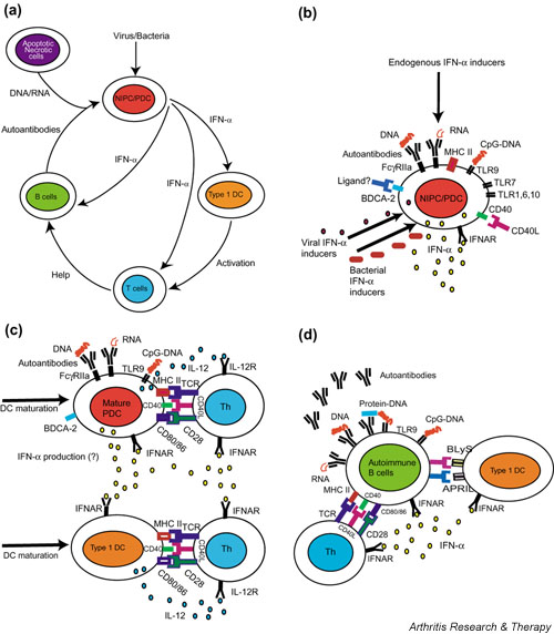
Patients with systemic lupus erythematosus (SLE) have ongoing interferon-alpha (IFN-alpha) production and serum IFN-alpha levels are correlated with both disease activity and severity. Recent studies of patients with SLE have demonstrated the presence of endogenous IFN-alpha inducers in such individuals, consisting of small immune complexes (ICs) containing IgG and DNA. These ICs act specifically on natural IFN-alpha-producing cells (NIPCs), often termed plasmacytoid dendritic cells (PDCs). Given the fact that the NIPC/PDC has a key role in both the innate and adaptive immune response, as well as the many immunoregulatory effects of IFN-alpha, these observations might be important for the understanding of the etiopathogenesis of SLE. In this review we briefly describe the biology of the type I IFN system, with emphasis on inducers, producing cells (especially NIPCs/PDCs), IFN-alpha actions and target immune cells that might be relevant in SLE. On the basis of this information and results from studies in SLE patients, we propose a hypothesis that explains how NIPCs/PDCs become activated and have a pivotal etiopathogenic role in SLE. This hypothesis also indicates new therapeutic targets in this autoimmune disease.


Figures

Figure 1
Figure 1
The central role of the type I interferon (IFN) system in the etiopathogenesis of systemic lupus erythematosus (SLE). (a) A schematic overview of IFN-α inducers and target cells. Initially, IFN-α is produced by the natural IFN-α producing cell (NIPC)/plasmacytoid dendritic cell (PDC) as a consequence of viral or bacterial infections. The IFN-α produced promotes DC1 development, T cell activation and autoantibody production by B cells. DNA or RNA and associated proteins, generated from apoptotic or necrotic cells, and autoantibodies form immune complexes (ICs) that act as endogenous IFN-α inducers and cause a prolonged IFN-α production. This IFN-α further stimulates the autoimmune response with more autoantibody production, IC formation, and co-stimulation of NIPCs/PDCs; finally, a vicious circle is created with an ongoing IFN-α production sustaining the autoimmune process. (b) Induction of IFN-α production in NIPCs/PDCs. Viruses, bacterial components, CpG-DNA and interferogenic ICs (IICs) can all trigger NIPCs/PDCs to produce IFN-α. FcγRIIa is necessary for the activation of NIPCs/PDCs by IICs. In addition, these cells express Toll-like receptor 9 (TLR9), mediating IFN-α synthesis induced by CpG-DNA, but the role of this receptor for the response to IICs is unknown. TLR7 activation by imiquimod also induces IFN-α production, but the function of TLR1, 6, and 10 in IFN-α production by NIPCs/PDCs is unknown. Ligation of CD40 enhances IFN-α synthesis and can also cause interleukin-12 (IL-12) production. In contrast, the ligation of blood dendritic cell antigen-2 (BDCA-2) by a monoclonal antibody inhibits the IFN-α production, but the natural ligand is unknown. IFNAR, IFN-α/β receptor. (c) Maturation of dendritic cells (DCs) and activation of T cells. The IFN-α produced induces the maturation of PDCs and the differentiation of monocytes to type 1 DCs; both cell types express the co-stimulatory molecules CD80 and CD86. These cells subsequently activate autoreactive T helper (Th) cells with specificity for processed antigens in IICs, for example. The cytokines IL-12 and IFN-α promote the Th1 response and prevent apoptosis in activated T cells. IL-12R, IL-12 receptor; TCR, T cell antigen receptor. (d) Production of autoantibodies by B cells. Activated Th cells provide help to B cells with reactivity to autoantigens in IICs, and these B cells are stimulated by IFN-α to prolonged survival and enhanced response to B cell antigen receptor (BCR) ligation. IFN-α also upregulates BLyS and APRIL ('a proliferation-inducing ligand') on DCs, which further promotes the B cell response and elicits CD40-independent Ig class switching and plasmacytoid differentiation. Autoantibody production is facilitated by the ability of DNA/RNA-containing autoantigens to activate B cells directly by simultaneous binding to BCR and TLR9. The autoantibodies produced bind to DNA and RNA and form more IICs, which trigger the continuous IFN-α production that is the fuel in the autoimmune process.

References

    1. Kelly JA, Moser KL, Harley JB. The genetics of systemic lupus erythematosus: putting the pieces together. Genes Immun. 2002;3(Suppl):S71–S85. - PubMed
    1. Tsao BP. An update on genetic studies of systemic lupus erythematosus. Curr Rheumatol Rep. 2002;4:359–367. - PubMed
    1. Grammer AC, Lipsky PE. CD154-CD40 interactions mediate differentiation to plasma cells in healthy individuals and persons with systemic lupus erythematosus. Arthritis Rheum. 2002;46:1417–1429. doi: 10.1002/art.10287. - DOI - PubMed
    1. Casciola-Rosen LA, Anhalt G, Rosen A. Autoantigens targeted in systemic lupus erythematosus are clustered in two populations of surface structures on apoptotic keratinocytes. J Exp Med. 1994;179:1317–1330. - PMC - PubMed
    1. Cocca BA, Cline AM, Radic MZ. Blebs and apoptotic bodies are B cell autoantigens. J Immunol. 2002;169:159–166. - PubMed

Publication types

MeSH terms