Comparative Study
. 2003 May;7(5 Pt 1):681-91.
doi: 10.1016/s1525-0016(03)00070-4.

Noninvasive imaging of lentiviral-mediated reporter gene expression in living mice

Affiliations
Comparative Study

Noninvasive imaging of lentiviral-mediated reporter gene expression in living mice

Abhijit De et al. Mol Ther. 2003 May.

Abstract

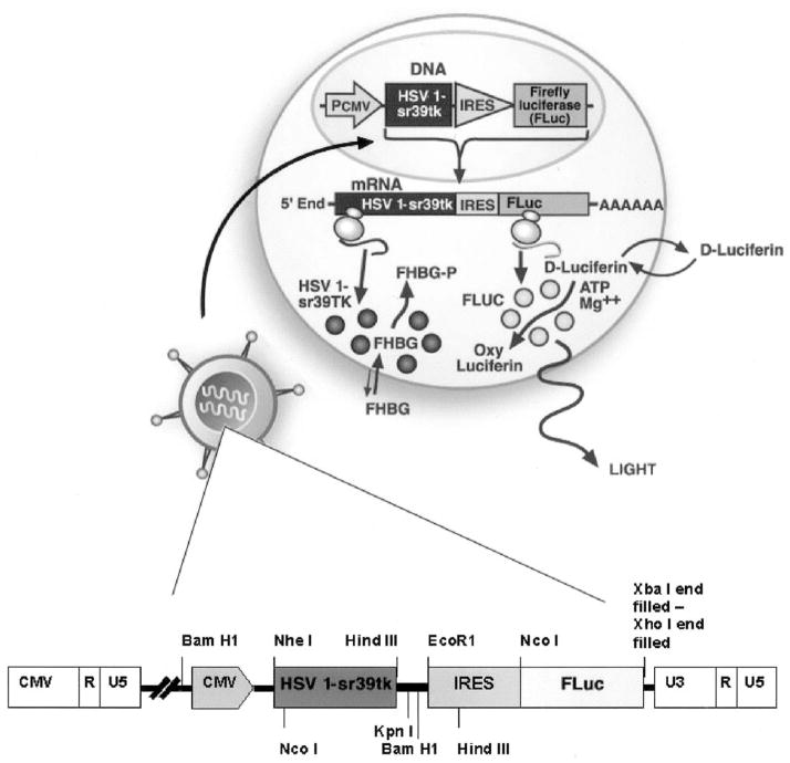
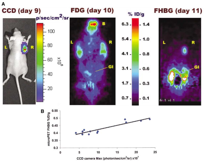
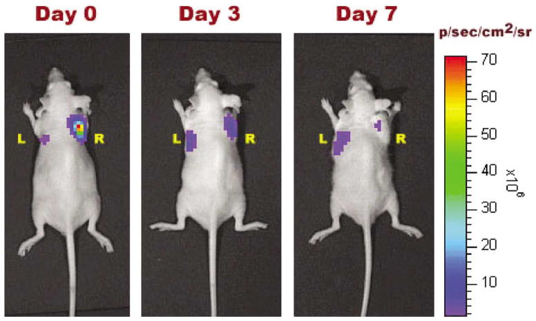
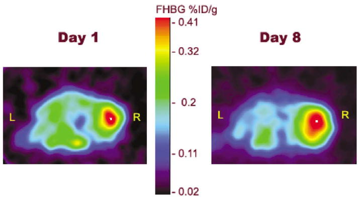
Lentiviral-mediated gene delivery holds significant promise for sustained gene expression within living systems. Vesicular stomatitis virus glycoprotein-pseudotyped human immunodeficiency virus type 1-based lentiviral vectors can be used to introduce transgenes in a broad spectrum of dividing as well as nondividing cells. In the current study, we construct a lentiviral vector carrying two reporter genes separated by an internal ribosomal entry site and utilize that virus in delivering both genes into neuroblastoma cells in cell culture and into cells implanted in living mice. We utilize two reporter genes, a mutant herpes simplex virus type 1 (HSV1) sr39tk as a reporter gene compatible with positron emission tomography (PET) and a bioluminescent optical reporter gene, firefly luciferase (Fluc), to image expression in living mice by an optical charge-coupled device (CCD) camera. By using this lentivirus, neuroblastoma (N2a) cells are stably transfected and a high correlation (R(2) = 0.91) between expressions of the two reporter genes in cell culture is established. Imaging of both reporter genes using microPET and optical CCD camera in living mice is feasible, with the optical approach being more sensitive, and a high correlation (R(2) = 0.86) between gene expressions is again observed in lentiviral-infected N2a tumor xenografts. Indirect imaging of HSV1-sr39tk suicide gene therapy utilizing Fluc is also feasible and can be detected with increased sensitivity by using the optical CCD. These preliminary results validate the use of lentiviral vectors carrying reporter genes for multimodality imaging of gene expression and should have many applications, including imaging of xenografts, metastasis, and cell trafficking as well as noninvasive monitoring of lentiviral-mediated gene delivery and expression.


Figures

FIG. 1
FIG. 1
Diagrammatic representation showing an IRES-based bicistronic lentivector used to link the activity of two reporter genes. The first gene (HSV1-sr39tk) is linked to the second gene (Fluc) by an EMCV IRES sequence. When this combination of genes is packaged within a lentivector for cell targeting, the virus delivers this DNA cassette to the host cell nucleus followed by a reverse transcription reaction. The two genes are integrated within the host genome irrespective of the cell division state and start producing messenger RNA as the host genome replicates. These mRNA molecules are translated to proteins within the host cytoplasm. By using FHBG as a PET reporter probe one can quantitatively image the HSV1-sr39TK activity within living subjects. The same is also true for the Fluc gene, in that by administration of D-Luciferin, a substrate for FLUC, light production can be imaged.
FIG. 2
FIG. 2
HSV1-sr39TK and FLUC enzyme activity in lentiviral (CS-CMVsr39tk-IFluc)-infected N2a cells. The TK activity is expressed as percentage conversion of 8-3H-PCV to its phosphorylated form per microgram protein per minute. The FLUC activity is expressed as RLU per microgram protein per second. Error bars represent SEM for triplicate measurements. (A) Relative HSV1-sr39TK enzyme activity in lentiviral CS-CMVsr39tk-I-Fluc-transduced N2a cells compared to parental N2a cells as a negative control. (B) Relative FLUC enzyme activity in lentiviral CS-CMVsr39tk-I-Fluc-transduced N2a cells compared with parental N2a cells as a negative control. (C) Correlation between HSV1-sr39TK (y axis) and FLUC (x axis) enzyme activity in lentiviral CS-CMVsr39tk-I-Fluc-transduced N2a cells as determined by assaying different numbers of infected cells. The correlation coefficient is R2 = 0.91.
FIG. 3
FIG. 3
Optical CCD and microPET imaging of a mouse implanted with N2a cells stably expressing HSV1-sr39tk and Fluc reporter genes. (A) Optical CCD image for Fluc expression on day 9, microPET FDG scan to check tumor viability on day 10, and microPET FHBG scan on day 11 for HSV1-sr39tk gene expression. Tumors were grown by injecting 5 × 10⁶ N2a cells transduced in culture by lentivirus (CS-CMVsr39tk-I-Fluc) (R) and control tumors an equal number of parental N2a cells (L). B, brain; GI, gastrointestinal tract; L, left tumor; R, right tumor. (B) Graph showing correlation of HSV1-sr39tk and Fluc gene expression in tumor xenografts. The correlation was established based on repeated scanning of four mice on days 8, 10, and 12 with both the microPET and the optical CCD camera. Individual mean FHBG %ID/g values versus maximum photons/s/cm2/sr values calculated from the regions of interest (ROI) on the respective scanned images of the tumor-bearing mice are plotted. Each of the 12 data points represents ROI values from bioluminescence (x axis) and microPET (y axis) images of the same mouse on the same day. The correlation coefficient is R2 = 0.86.
FIG. 4
FIG. 4
Optical CCD images of tumor xenografts showing Fluc gene expression over time. Tumors were implanted sc with 5 × 10⁵ lentiviral (CS-CMVsr39tk-I-Fluc)-infected N2a cells on the right shoulder (R) and same number of parental N2a cells on the left shoulder (L) on day 0. The mouse was repeatedly scanned with the CCD on days 0, 4, 7, 11, and 14 as the tumors grew. The highest expression was observed on day 11, after which internal necrosis within the tumors likely resulted in lowered intensity of FLUC signal on day 14. All images shown are the visible light image superimposed on the optical CCD bioluminescence images.
FIG. 5
FIG. 5
Monitoring of HSV1-sr39tk suicide gene therapy indirectly by Fluc reporter gene expression in a tumor xenograft model. In a set of six mice, control tumors (L) with N2a cells infected with lentivirus CS-CMVFluc and experimental tumors (R) with N2a cells infected with lentivirus CS-CMVsr39tk-I-Fluc were implanted with 2 ×10⁶ cells. Tumors were allowed to grow for 6 days before GCV treatment was started (day 0). GCV was administered daily at a dose of 25 mg/mouse/day. Follow-up scans with D-Luciferin on days 3 and 7 of GCV treatment showed significant decrease in the FLUC signal at the site of the experimental tumor (R), whereas the control tumor (L) continued to grow with increasing light signal.
FIG. 6
FIG. 6
Optical CCD imaging of Fluc gene expression over 7 days in a living mouse carrying xenografts. On the left, tumors were implanted sc with parental N2a cells on both shoulders and allowed to grow to a palpable size of ~5 mm diameter before lentivirus (CS-CMVsr39tk-I-Fluc) was injected directly into the right tumor (R) only once. The animals were scanned for Fluc expression on days 2, 4, and 7 after virus injection. On the right, tumors were implanted sc with parental N2a cells on the left shoulder (L) and with lentiviral (CS-CMVsr39tk-I-Fluc)-infected N2a cells on the right shoulder (R). These animals were scanned on days 0 (2 h after cell implantation), 4, and 7.
FIG. 7
FIG. 7
FHBG microPET images of transverse sections of a mouse on days 1 and 8 with HSV1-sr39tk gene expression in a tumor xenograft. N2a parental cells were implanted sc on both shoulders and allowed to grow to a palpable size of ~5 mm diameter before lentivirus (CS-CMVsr39tk-I-Fluc) was injected directly into the right tumor (R) only once. L indicates left tumor.

References

    1. Verma IM, Somia N. Gene therapy—Promises, problems and prospects. Nature. 1997;389:239–242. - PubMed
    1. Lewis PF, Emerman M. Passage through mitosis is required for oncoret-roviruses but not for the human immunodeficiency virus. J Virol. 1994;68:510–516. - PMC - PubMed
    1. Naldini L, et al. Efficient transfer, integration, and sustained long-term expression of the transgene in adult rat brains injected with a lentiviral vector. Proc Natl Acad Sci USA. 1996;93:11382–11388. - PMC - PubMed
    1. Miyoshi H, et al. Development of a self-inactivating lentivirus vector. J Virol. 1998;72:8150–8157. - PMC - PubMed
    1. Yu D, et al. Prostate-specific targeting using PSA promoter-based lentiviral vectors. Cancer Gene Ther. 2001;8:628–635. - PubMed

Publication types

MeSH terms