

EPR spectroscopy shows a microtubule-dependent conformational change in the kinesin switch 1 domain
- PMID: 12719248
- PMCID: PMC1302879
- DOI: 10.1016/S0006-3495(03)70043-5
EPR spectroscopy shows a microtubule-dependent conformational change in the kinesin switch 1 domain
Abstract
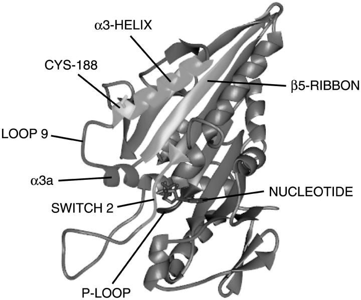
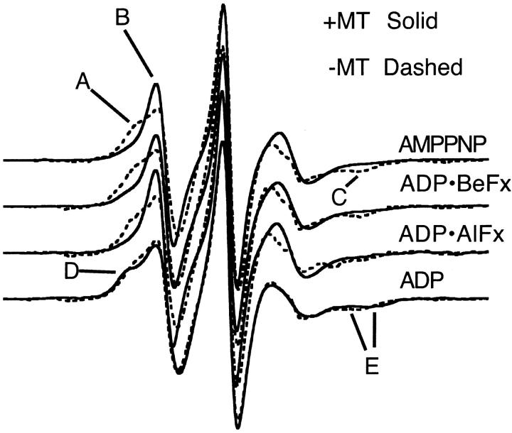
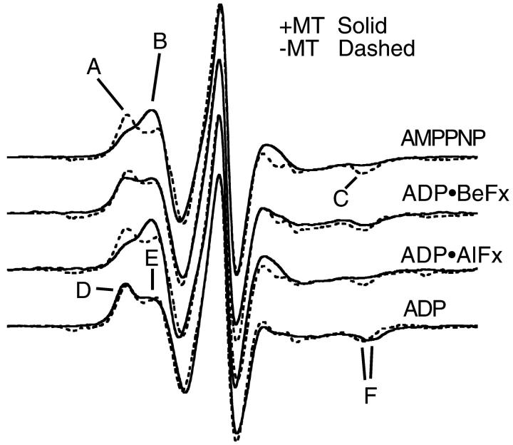
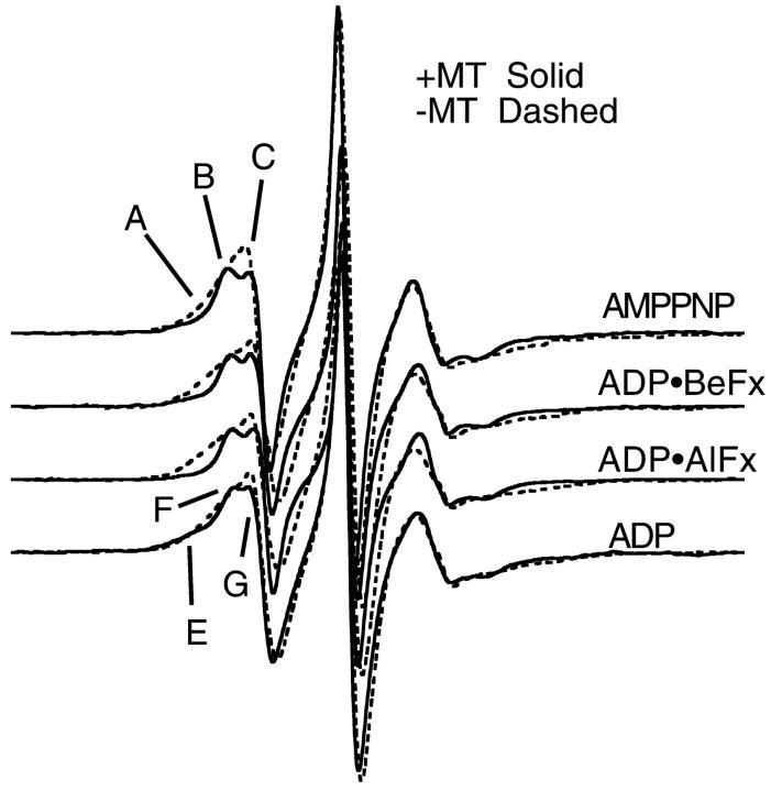
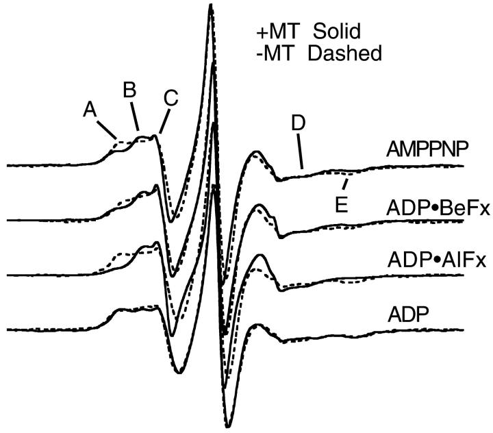
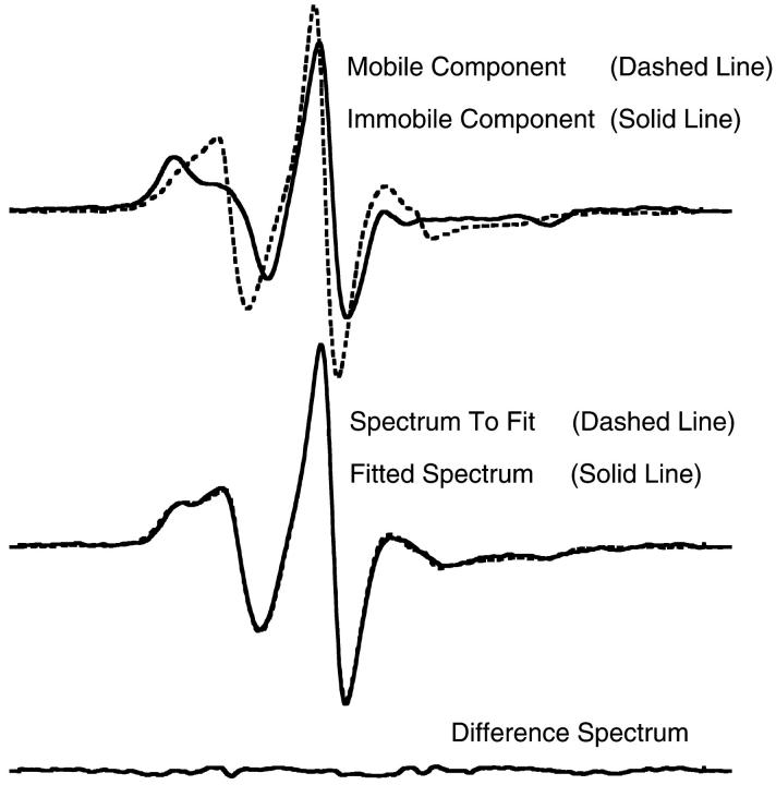
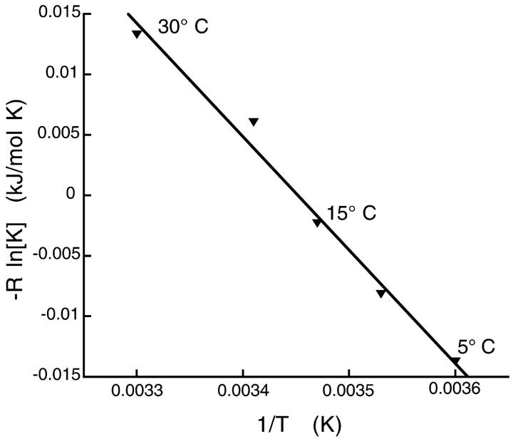
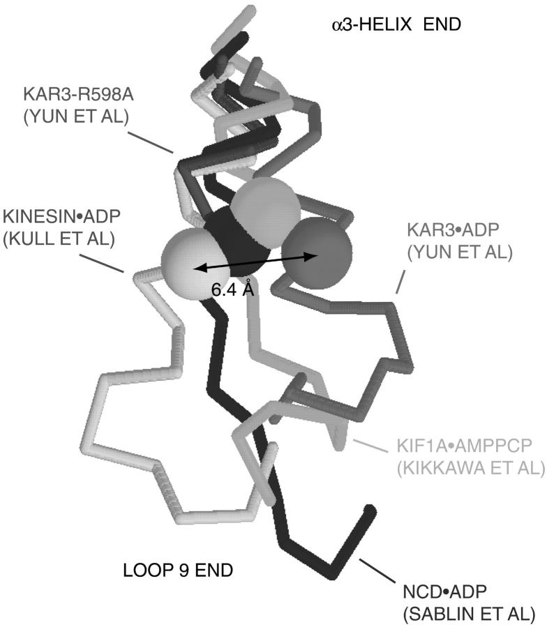
We have used site-directed spin-labeling and electron paramagnetic resonance spectroscopy to monitor a conformational change at the nucleotide site of kinesin. Cys-lite kinesin (K349 monomer) with the mutation S188C was spin labeled with MSL or MTSL. This residue is at the junction between the switch 1 region (which is a structure known to be sensitive to bound nucleotide in the G-proteins) and the alpha3-helix, adjacent to the nucleotide site. The spectra showed two or more components of mobility, which were independent of nucleotide in the absence of microtubules (MTs). The spectra of both labels showed a change of mobility upon binding to MTs. A more mobile spectral component became enhanced for all triphosphate analogs examined, AMPPNP, ADP.AlFx, or ADP.BeFx, in the presence of MTs, although the magnitude of the new component and the degree of mobility varied with nucleotide analog. The ADP state showed a much-reduced spectral change with a small shift to the more immobilized component in the presence of MTs. For kinesin.ADP.MT, a van't Hoff plot gave DeltaH degrees = -96 kJ/mol implying that the conformational change was extensive. We conclude there is a conformational change in the switch 1-alpha3-helix domain when kinesin binds to MTs.
Figures
Similar articles
-
Nucleotide-dependent displacement and dynamics of the α-1 helix in kinesin revealed by site-directed spin labeling EPR.Biochem Biophys Res Commun. 2014 Jan 17;443(3):911-6. doi: 10.1016/j.bbrc.2013.12.063. Epub 2013 Dec 19. Biochem Biophys Res Commun. 2014. PMID: 24361895
-
Multiple conformations of the nucleotide site of Kinesin family motors in the triphosphate state.J Mol Biol. 2011 May 13;408(4):628-42. doi: 10.1016/j.jmb.2011.01.001. Epub 2011 Jan 26. J Mol Biol. 2011. PMID: 21277856 Free PMC article.
-
Thermodynamic properties of the kinesin neck-region docking to the catalytic core.Biophys J. 2003 Mar;84(3):1844-54. doi: 10.1016/S0006-3495(03)74992-3. Biophys J. 2003. PMID: 12609886 Free PMC article.
-
Kinesin: switch I & II and the motor mechanism.J Cell Sci. 2002 Jan 1;115(Pt 1):15-23. doi: 10.1242/jcs.115.1.15. J Cell Sci. 2002. PMID: 11801720 Review.
-
A look into kinesin's powerhouse.FEBS Lett. 2001 Nov 23;508(3):291-4. doi: 10.1016/s0014-5793(01)03064-2. FEBS Lett. 2001. PMID: 11728437 Review.
Cited by
-
Dynamics of the nucleotide pocket of myosin measured by spin-labeled nucleotides.Biophys J. 2007 Jan 1;92(1):172-84. doi: 10.1529/biophysj.106.090035. Epub 2006 Oct 6. Biophys J. 2007. PMID: 17028139 Free PMC article.
-
Shaft Function of Kinesin-1's α4 Helix in the Processive Movement.Cell Mol Bioeng. 2019 Jun 25;12(4):345-354. doi: 10.1007/s12195-019-00581-4. eCollection 2019 Aug. Cell Mol Bioeng. 2019. PMID: 31719918 Free PMC article.
-
Microtubule-kinesin interface mutants reveal a site critical for communication.Biochemistry. 2004 Mar 16;43(10):2792-803. doi: 10.1021/bi035830e. Biochemistry. 2004. PMID: 15005614 Free PMC article.
-
The Kinesin-1 tail conformationally restricts the nucleotide pocket.Biophys J. 2009 Apr 8;96(7):2799-807. doi: 10.1016/j.bpj.2008.11.069. Biophys J. 2009. PMID: 19348763 Free PMC article.
-
Anchor Effect of Interactions Between Kinesin's Nucleotide-Binding Pocket and Microtubule.Cell Mol Bioeng. 2017 Feb 15;10(2):162-173. doi: 10.1007/s12195-017-0477-8. eCollection 2017 Apr. Cell Mol Bioeng. 2017. PMID: 31719858 Free PMC article.
References
-
- Baumann, B. A., B. D. Hambly, K. Hideg, and P. G. Fajer. 2001. The regulatory domain of the myosin head behaves as a rigid lever. Biochemistry. 40:7868–7873. - PubMed
-
- Boriack-Sjodin, P. A., S. M. Margarit, D. Bar-Sagi, and J. Kuriyan. 1998. The structural basis of the activation of Ras by Sos. Nature. 394:337–343. - PubMed
-
- Bradford, M. M. 1976. A rapid and sensitive method for the quantitation of microgram quantities of protein utilizing the principle of protein-dye binding. Anal. Biochem. 72:248–254. - PubMed
MeSH terms
Substances
Grants and funding
LinkOut - more resources
Full Text Sources
