

Human aortic valve calcification is associated with an osteoblast phenotype
- PMID: 12719282
- PMCID: PMC3922288
- DOI: 10.1161/01.CIR.0000070591.21548.69
Human aortic valve calcification is associated with an osteoblast phenotype
Abstract
Background: Calcific aortic stenosis is the third most common cardiovascular disease in the United States. We hypothesized that the mechanism for aortic valve calcification is similar to skeletal bone formation and that this process is mediated by an osteoblast-like phenotype.
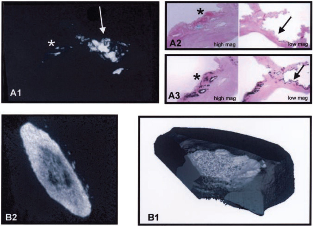
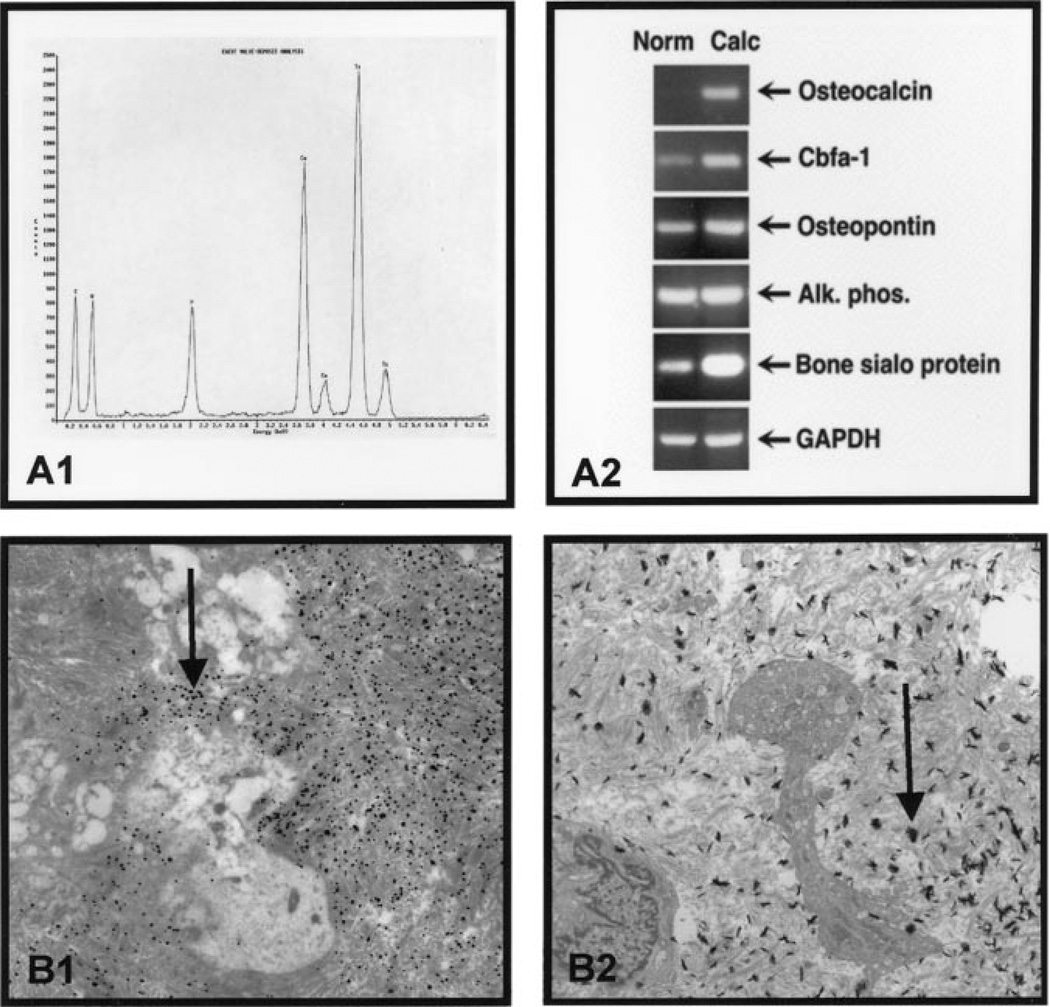
Methods and results: To test this hypothesis, we examined calcified human aortic valves replaced at surgery (n=22) and normal human valves (n=20) removed at time of cardiac transplantation. Contact microradiography and micro-computerized tomography were used to assess the 2-dimensional and 3-dimensional extent of mineralization. Mineralization borders were identified with von Kossa and Goldner's stains. Electron microscopy and energy-dispersive spectroscopy were performed for identification of bone ultrastructure and CaPO4 composition. To analyze for the osteoblast and bone markers, reverse transcriptase-polymerase chain reaction was performed on calcified versus normal human valves for osteopontin, bone sialoprotein, osteocalcin, alkaline phosphatase, and the osteoblast-specific transcription factor Cbfa1. Microradiography and micro-computerized tomography confirmed the presence of calcification in the valve. Special stains for hydroxyapatite and CaPO4 were positive in calcification margins. Electron microscopy identified mineralization, whereas energy-dispersive spectroscopy confirmed the presence of elemental CaPO4. Reverse transcriptase-polymerase chain reaction revealed increased mRNA levels of osteopontin, bone sialoprotein, osteocalcin, and Cbfa1 in the calcified valves. There was no change in alkaline phosphatase mRNA level but an increase in the protein expression in the diseased valves.
Conclusions: These findings support the concept that aortic valve calcification is not a random degenerative process but an active regulated process associated with an osteoblast-like phenotype.
Figures
Comment in
-
Human aortic valve calcification.Circulation. 2003 Dec 9;108(23):e163; author reply e163. doi: 10.1161/01.CIR.0000102948.81404.D8. Circulation. 2003. PMID: 14662698 No abstract available.
References
-
- Lindroos M, Kupari M, Heikkila J, et al. Prevalence of aortic valve abnormalities in the elderly: an echocardiographic study of a random population sample. J Am Coll Cardiol. 1993;21:1220–1225. - PubMed
-
- Ross J, Braunwald E. Aortic stenosis. Circulation. 1968;381(suppl):61–67. - PubMed
-
- Mohler ER, Gannon F, Reynolds C, et al. Bone formation and inflammation in cardiac valves. Circulation. 2001;103:1522–1528. - PubMed
-
- O’Brien KD, Kuusisto J, Reichenbach DD, et al. Osteopontin is expressed in human aortic valvular lesions. Circulation. 1995;92:2163–2168. - PubMed
-
- Mohler ER, Chawla MK, Chang AW, et al. Identification and characterization of calcifying valve cells from human and canine aortic valves. J Heart Valve Dis. 1999;8:254–260. - PubMed
MeSH terms
Substances
Grants and funding
LinkOut - more resources
Full Text Sources
Other Literature Sources
Medical
Research Materials
