. 2003 Apr 28;161(2):249-55.
doi: 10.1083/jcb.200208039.

Helicobacter pylori CagA protein targets the c-Met receptor and enhances the motogenic response

Affiliations

Helicobacter pylori CagA protein targets the c-Met receptor and enhances the motogenic response

Yuri Churin et al. J Cell Biol. .

Abstract

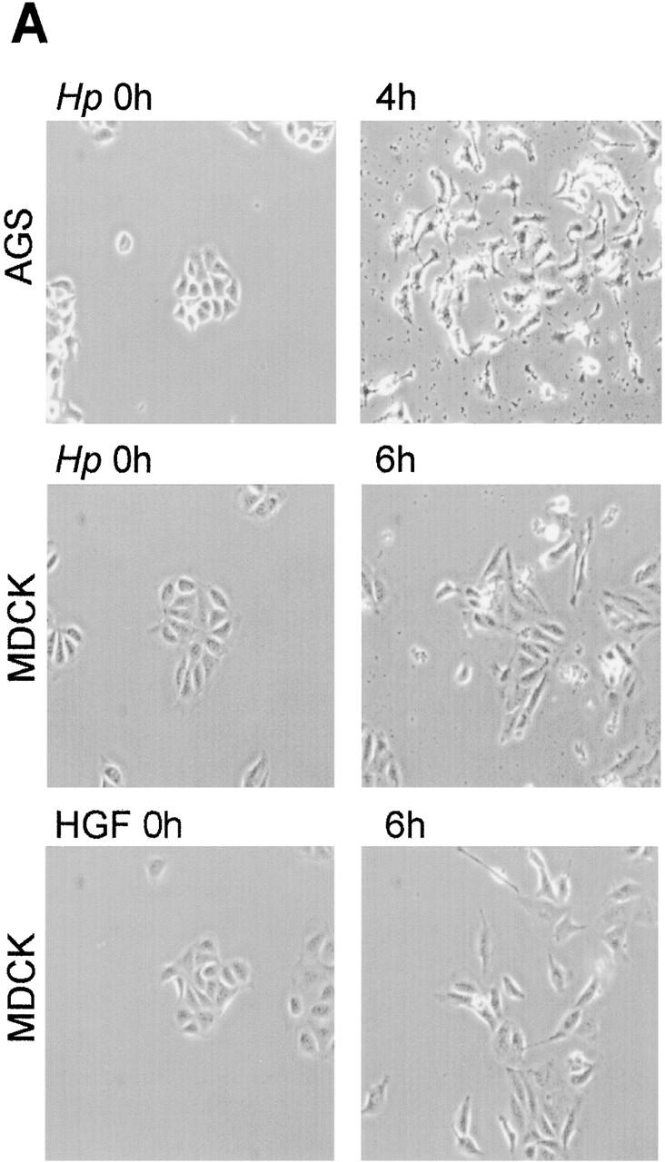
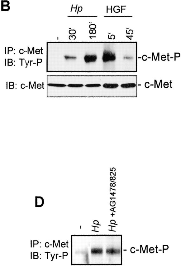
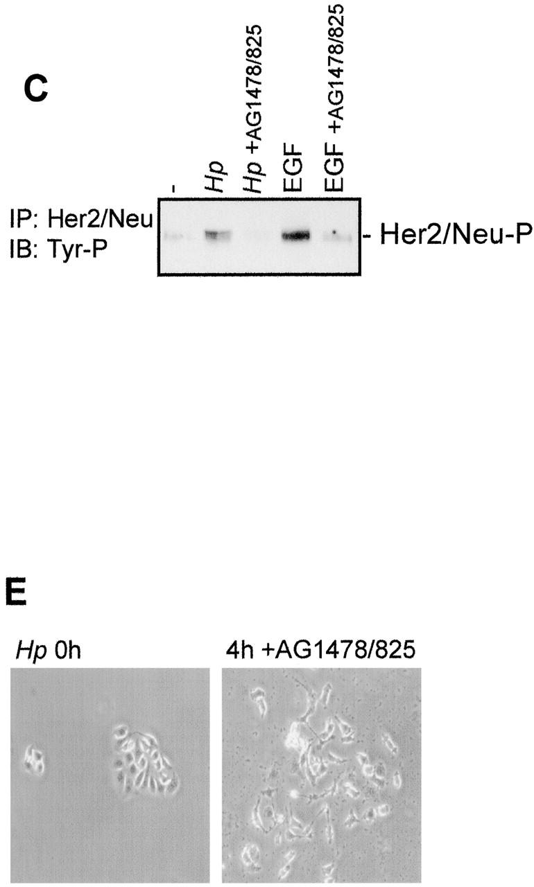
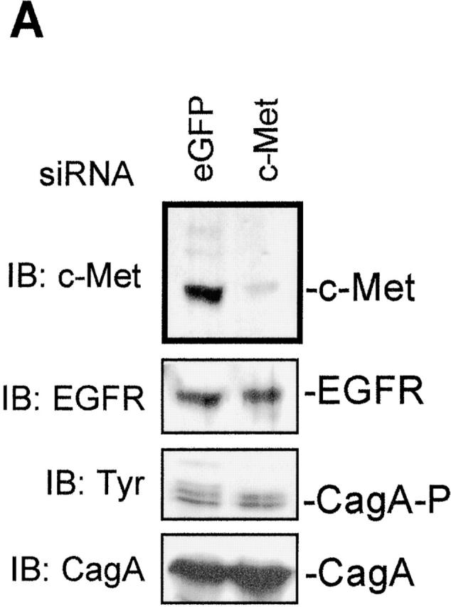
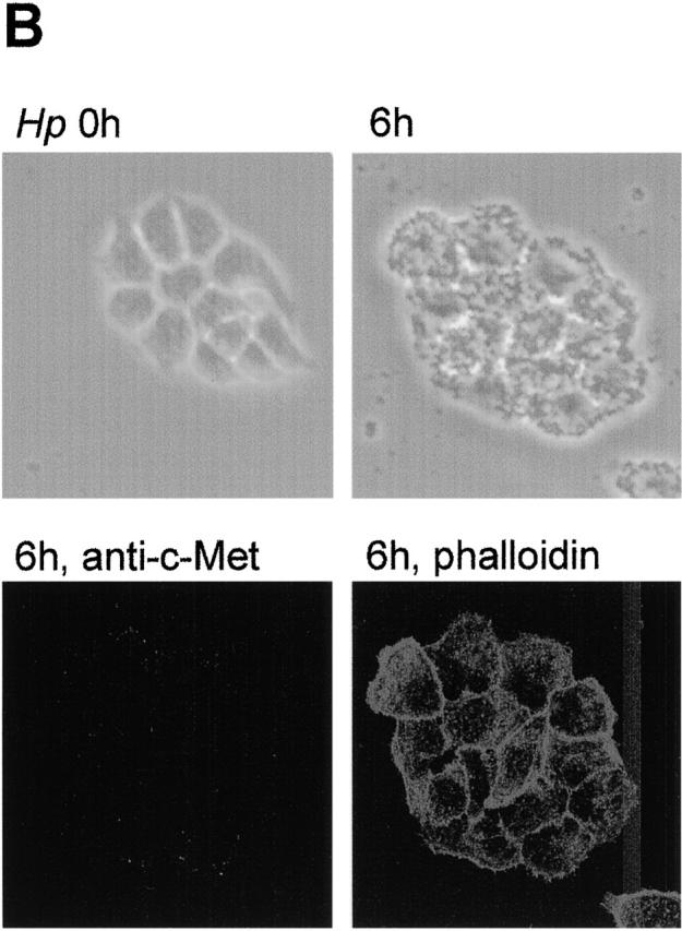
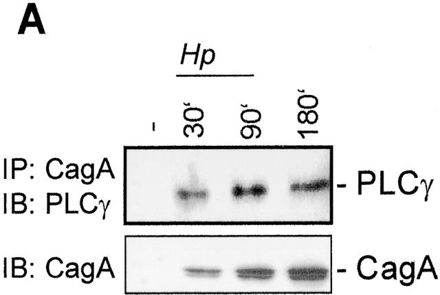
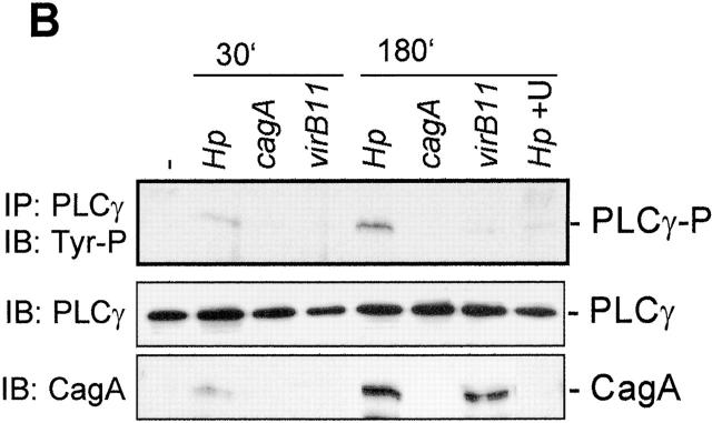
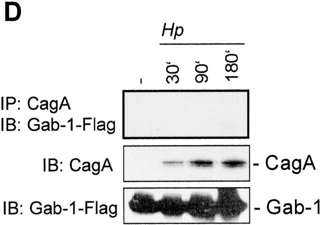
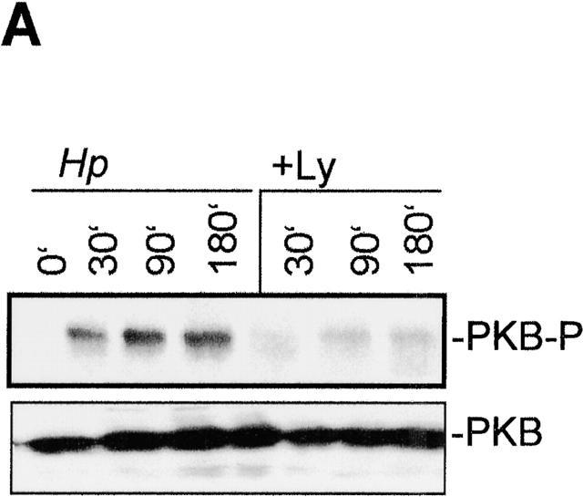
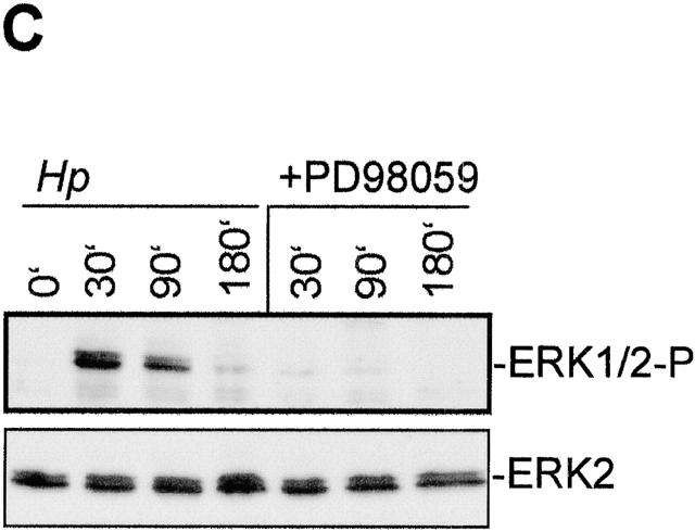
Infection with the human microbial pathogen Helicobacter pylori is assumed to lead to invasive gastric cancer. We find that H. pylori activates the hepatocyte growth factor/scatter factor receptor c-Met, which is involved in invasive growth of tumor cells. The H. pylori effector protein CagA intracellularly targets the c-Met receptor and promotes cellular processes leading to a forceful motogenic response. CagA could represent a bacterial adaptor protein that associates with phospholipase Cgamma but not Grb2-associated binder 1 or growth factor receptor-bound protein 2. The H. pylori-induced motogenic response is suppressed and blocked by the inhibition of PLCgamma and of MAPK, respectively. Thus, upon translocation, CagA modulates cellular functions by deregulating c-Met receptor signaling. The activation of the motogenic response in H. pylori-infected epithelial cells suggests that CagA could be involved in tumor progression.


Figures

Figure 1.
Figure 1.
H. pylori activates c-Met receptor tyrosine kinase and induces the motogenic response. (A) H. pylori infection induces motility of AGS and MDCK cells. AGS and MDCK cells were infected with H. pylori, or MDCK cells were treated with 50 U/ml HGF. Phase-contrast microscopy was performed at the indicated time points. (B) H. pylori activates the c-Met receptor in AGS cells. AGS cells were infected with H. pylori or treated with HGF. c-Met was immunoprecipitated from lysates prepared at the indicated time points. Immunoprecipitates (IP) were subjected to SDS-PAGE and immunoblot (IB) analysis with antiphosphotyrosine (top) or anti–c-Met (bottom) antibodies. (C) H. pylori infection activates HER2/Neu. AGS cells were pretreated with or without AG1478 and AG825, and either infected with H. pylori for 90 min or treated with 10 ng/ml EGF for 5 min. Cell lysates were prepared, and HER2/Neu was immunoprecipitated and subjected to Western blot analysis using antiphosphotyrosine antibody. (D) AG1478 and AG825 have no effect on c-Met activation. AGS cells were pretreated with or without AG1478 and AG825 and infected with H. pylori for 180 min, and c-Met was immunoprecipitated and subjected to Western blot analysis using antiphosphotyrosine antibody. (E) The inhibitors of EGFR and HER2/Neu had no effect on the motility of AGS cells. AGS cells were treated with the inhibitors of EGFR (AG1478) and HER2/Neu (AG825) and infected with H. pylori. Phase-contrast microscopy was performed 4 h after infection.
Figure 1.
Figure 1.
H. pylori activates c-Met receptor tyrosine kinase and induces the motogenic response. (A) H. pylori infection induces motility of AGS and MDCK cells. AGS and MDCK cells were infected with H. pylori, or MDCK cells were treated with 50 U/ml HGF. Phase-contrast microscopy was performed at the indicated time points. (B) H. pylori activates the c-Met receptor in AGS cells. AGS cells were infected with H. pylori or treated with HGF. c-Met was immunoprecipitated from lysates prepared at the indicated time points. Immunoprecipitates (IP) were subjected to SDS-PAGE and immunoblot (IB) analysis with antiphosphotyrosine (top) or anti–c-Met (bottom) antibodies. (C) H. pylori infection activates HER2/Neu. AGS cells were pretreated with or without AG1478 and AG825, and either infected with H. pylori for 90 min or treated with 10 ng/ml EGF for 5 min. Cell lysates were prepared, and HER2/Neu was immunoprecipitated and subjected to Western blot analysis using antiphosphotyrosine antibody. (D) AG1478 and AG825 have no effect on c-Met activation. AGS cells were pretreated with or without AG1478 and AG825 and infected with H. pylori for 180 min, and c-Met was immunoprecipitated and subjected to Western blot analysis using antiphosphotyrosine antibody. (E) The inhibitors of EGFR and HER2/Neu had no effect on the motility of AGS cells. AGS cells were treated with the inhibitors of EGFR (AG1478) and HER2/Neu (AG825) and infected with H. pylori. Phase-contrast microscopy was performed 4 h after infection.
Figure 2.
Figure 2.
c-Met receptor expression is essential for H. pylori– induced motogenic response in epithelial cells. HeLa cells were transfected with siRNA to c-Met or with siRNA to EGFP (as a control for the effect of transfection). After culturing for 72 h, cells were infected with the H. pylori strain P1 for 6 h. The siRNA to c-Met efficiently silenced c-Met receptor expression analyzed in a Western blot (A, top). Silencing of c-Met expression had no effect on EGFR expression (second panel) and phosphorylation (third panel) of translocated CagA protein (bottom). (B) Cells were transfected with c-Met siRNA and infected with the wild-type H. pylori strain P1. Cells are shown by phase-contrast microscopy (top two panels) or stained with immunofluorescence using c-Met antibody. Actin filaments were visualized with rhodamine-conjugated phalloidin. (C) HeLa cells transfected with c-Met siRNA are resistant to the induction of the motogenic response by H. pylori. Phase-contrast of cells transfected with siRNA to c-Met or siRNA to EGFP.
Figure 2.
Figure 2.
c-Met receptor expression is essential for H. pylori– induced motogenic response in epithelial cells. HeLa cells were transfected with siRNA to c-Met or with siRNA to EGFP (as a control for the effect of transfection). After culturing for 72 h, cells were infected with the H. pylori strain P1 for 6 h. The siRNA to c-Met efficiently silenced c-Met receptor expression analyzed in a Western blot (A, top). Silencing of c-Met expression had no effect on EGFR expression (second panel) and phosphorylation (third panel) of translocated CagA protein (bottom). (B) Cells were transfected with c-Met siRNA and infected with the wild-type H. pylori strain P1. Cells are shown by phase-contrast microscopy (top two panels) or stained with immunofluorescence using c-Met antibody. Actin filaments were visualized with rhodamine-conjugated phalloidin. (C) HeLa cells transfected with c-Met siRNA are resistant to the induction of the motogenic response by H. pylori. Phase-contrast of cells transfected with siRNA to c-Met or siRNA to EGFP.
Figure 3.
Figure 3.
CagA interacts with the c-Met tyrosine kinase receptor and enhances the motogenic response of AGS cells to H. pylori infection. (A and B) AGS cells were infected with the wild-type H. pylori strain or isogenic mutant strains cagA and virB11. Phase-contrast microscopy was performed at the indicated time points. (B) Different H. pylori strains activate c-Met. Cells were harvested at the indicated time points after infection. c-Met was immunoprecipitated (IP) with anti–c-Met antibody and analyzed by immunoblotting (IB) using antiphosphotyrosine antibody. (C and D) CagA interacts with the c-Met receptor. AGS cells were infected with H. pylori. Cell lysates were prepared at the indicated time points. c-Met (C) or CagA (D) were immunoprecipitated with the corresponding specific antibodies and subjected to immunoblot analysis using anti-CagA (C) and anti–c-Met (D) antibodies. (E) CagA–c-Met interaction depends on c-Met tyrosine phosphorylation. AGS cells were transiently transfected with plasmids expressing either HA-tagged wild-type CagA (CagA) or HA-tagged phosphorylation-resistant CagA (CagAΔP) and were treated with HGF for 5 min. CagA was precipitated with anti-HA antibody, and immunoprecipitates were analyzed by Western blot analysis using anti–c-Met (top), antiphosphotyrosine (middle) antibodies, and anti–c-Met (bottom) antibodies.
Figure 3.
Figure 3.
CagA interacts with the c-Met tyrosine kinase receptor and enhances the motogenic response of AGS cells to H. pylori infection. (A and B) AGS cells were infected with the wild-type H. pylori strain or isogenic mutant strains cagA and virB11. Phase-contrast microscopy was performed at the indicated time points. (B) Different H. pylori strains activate c-Met. Cells were harvested at the indicated time points after infection. c-Met was immunoprecipitated (IP) with anti–c-Met antibody and analyzed by immunoblotting (IB) using antiphosphotyrosine antibody. (C and D) CagA interacts with the c-Met receptor. AGS cells were infected with H. pylori. Cell lysates were prepared at the indicated time points. c-Met (C) or CagA (D) were immunoprecipitated with the corresponding specific antibodies and subjected to immunoblot analysis using anti-CagA (C) and anti–c-Met (D) antibodies. (E) CagA–c-Met interaction depends on c-Met tyrosine phosphorylation. AGS cells were transiently transfected with plasmids expressing either HA-tagged wild-type CagA (CagA) or HA-tagged phosphorylation-resistant CagA (CagAΔP) and were treated with HGF for 5 min. CagA was precipitated with anti-HA antibody, and immunoprecipitates were analyzed by Western blot analysis using anti–c-Met (top), antiphosphotyrosine (middle) antibodies, and anti–c-Met (bottom) antibodies.
Figure 4.
Figure 4.
H. pylori–activated PLCγ interacts with CagA and is required for the motogenic response of AGS cells. (A) PLCγ interacts with CagA. CagA was immunoprecipitated (IP) from AGS cell lysates prepared at the indicated time points after infection. Immunoblot (IB) analysis of immunoprecipitates was performed using anti-PLCγ antibody. (B) H. pylori induces PLCγ phosphorylation. AGS cells were infected with H. pylori wild type or cagA and virB11 strains, and PLCγ immunoprecipitates were tested by Western blot analysis using antiphosphotyrosine antibody. (C) PLCγ inhibitor drastically reduces the motogenic response of AGS cells to H. pylori infection. AGS cells were pretreated with the PLCγ inhibitor U73122 and infected with H. pylori. Phase-contrast microscopy was performed at 4 h after infection. (D) CagA fails to interact with large adaptor protein Gab1. AGS cells were transiently transfected with the plasmid-expressed Flag-tagged Gab1 and infected with H. pylori. CagA immunoprecipitates were analyzed by immunoblotting with anti-Flag antibody.
Figure 4.
Figure 4.
H. pylori–activated PLCγ interacts with CagA and is required for the motogenic response of AGS cells. (A) PLCγ interacts with CagA. CagA was immunoprecipitated (IP) from AGS cell lysates prepared at the indicated time points after infection. Immunoblot (IB) analysis of immunoprecipitates was performed using anti-PLCγ antibody. (B) H. pylori induces PLCγ phosphorylation. AGS cells were infected with H. pylori wild type or cagA and virB11 strains, and PLCγ immunoprecipitates were tested by Western blot analysis using antiphosphotyrosine antibody. (C) PLCγ inhibitor drastically reduces the motogenic response of AGS cells to H. pylori infection. AGS cells were pretreated with the PLCγ inhibitor U73122 and infected with H. pylori. Phase-contrast microscopy was performed at 4 h after infection. (D) CagA fails to interact with large adaptor protein Gab1. AGS cells were transiently transfected with the plasmid-expressed Flag-tagged Gab1 and infected with H. pylori. CagA immunoprecipitates were analyzed by immunoblotting with anti-Flag antibody.
Figure 5.
Figure 5.
H. pylori–induced motogenic response requires ERK activity. (A) H. pylori induces PI3-K–dependent PKB phosphorylation. AGS cells were pretreated with inhibitor of PI3-K (Ly) and infected with H. pylori. Total cell lysates were prepared at the indicated time points after infection and analyzed by Western blot analysis using phospho-PKB (Ser473) antibody. (B) Inhibitor of PI3-K fails to suppress the motogenic response of AGS cells after H. pylori infection. AGS cells were pretreated with Ly294002 and infected with H. pylori. Phase-contrast microscopy was performed at 4 h after infection. (C) Pretreatment of AGS cells with MAPK/extracellular regulated kinase (MEK) inhibitor PD98059 inhibits ERK1/2 activation by H. pylori. AGS cells were pretreated with or without PD98059 and infected with H. pylori. Total cell lysates were prepared at the indicated time points after infection and analyzed by Western blot analysis using phospho-p44/42 MAPK antibody. (D) Treatment with the MEK inhibitor PD98059 blocks the motogenic response in AGS cells. AGS cells were pretreated with the MEK inhibitor PD98059 and infected with H. pylori. Phase-contrast microscopy was performed 4 h after infection.
Figure 5.
Figure 5.
H. pylori–induced motogenic response requires ERK activity. (A) H. pylori induces PI3-K–dependent PKB phosphorylation. AGS cells were pretreated with inhibitor of PI3-K (Ly) and infected with H. pylori. Total cell lysates were prepared at the indicated time points after infection and analyzed by Western blot analysis using phospho-PKB (Ser473) antibody. (B) Inhibitor of PI3-K fails to suppress the motogenic response of AGS cells after H. pylori infection. AGS cells were pretreated with Ly294002 and infected with H. pylori. Phase-contrast microscopy was performed at 4 h after infection. (C) Pretreatment of AGS cells with MAPK/extracellular regulated kinase (MEK) inhibitor PD98059 inhibits ERK1/2 activation by H. pylori. AGS cells were pretreated with or without PD98059 and infected with H. pylori. Total cell lysates were prepared at the indicated time points after infection and analyzed by Western blot analysis using phospho-p44/42 MAPK antibody. (D) Treatment with the MEK inhibitor PD98059 blocks the motogenic response in AGS cells. AGS cells were pretreated with the MEK inhibitor PD98059 and infected with H. pylori. Phase-contrast microscopy was performed 4 h after infection.

References

    1. Asahi, M., T. Azuma, S. Ito, Y. Ito, H. Suto, Y. Nagai, M. Tsubokawa, Y. Tohyama, S. Maeda, M. Omata, et al. 2000. Helicobacter pylori CagA protein can be tyrosine phosphorylated in gastric epithelial cells. J. Exp. Med. 191:593–602. - PMC - PubMed
    1. Backert, S., E. Ziska, V. Brinkmann, U. Zimny-Arndt, A. Fauconnier, P.R. Jungblut, M. Naumann, and T.F. Meyer. 2000. Translocation of the Helicobacter pylori CagA protein in gastric epithelial cells by a type IV secretion apparatus. Cell Microbiol. 2:155–164. - PubMed
    1. Bardelli, A., P. Longati, D. Gramaglia, C. Basilico, L. Tamagnone, S. Giordano, D. Ballinari, P. Michieli, and P.M. Comoglio. 1998. Uncoupling signal transducers from oncogenic MET mutants abrogates cell transformation and inhibits invasive growth. Proc. Natl. Acad. Sci. USA. 95:14379–14383. - PMC - PubMed
    1. Carpenter, G., and Q. Ji. 1999. Phospholipase C-gamma as a signal-transducing element. Exp. Cell Res. 253:15–24. - PubMed
    1. Chen, P., J.E. Murphy-Ullrich, and A. Wells. 1996. A role for gelsolin in actuating epidermal growth factor receptor–mediated cell motility. J. Cell Biol. 134:689–698. - PMC - PubMed

Publication types

MeSH terms