Expression of a targeted lambda 1 light chain gene is developmentally regulated and independent of Ig kappa rearrangements
- PMID: 12719477
- PMCID: PMC2193966
- DOI: 10.1084/jem.20030402
Expression of a targeted lambda 1 light chain gene is developmentally regulated and independent of Ig kappa rearrangements
Erratum in
- J Exp Med. 2003 May 19;1399
Abstract
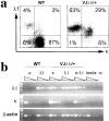
Immunoglobulin light chain (IgL) rearrangements occur more frequently at Ig kappa than at Ig lambda. Previous results suggested that the unrearranged Ig kappa locus negatively regulates Ig lambda transcription and/or rearrangement. Here, we demonstrate that expression of a VJ lambda 1-joint inserted into its physiological position in the Ig lambda locus is independent of Ig kappa rearrangements. Expression of the inserted VJ lambda 1 gene segment is developmentally controlled like that of a VJ kappa-joint inserted into the Ig kappa locus and furthermore coincides developmentally with the occurrence of Ig kappa rearrangements in wild-type mice. We conclude that developmentally controlled transcription of a gene rearrangement in the Ig lambda locus occurs in the presence of an unrearranged Ig kappa locus and is therefore not negatively regulated by the latter. Our data also indicate light chain editing in approximately 30% of lambda 1 expressing B cell progenitors.
Figures


Similar articles
-
Gene targeting in the Ig kappa locus: efficient generation of lambda chain-expressing B cells, independent of gene rearrangements in Ig kappa.EMBO J. 1993 Mar;12(3):811-20. doi: 10.1002/j.1460-2075.1993.tb05721.x. EMBO J. 1993. PMID: 8458339 Free PMC article.
-
Light chain editing in kappa-deficient animals: a potential mechanism of B cell tolerance.J Exp Med. 1994 Nov 1;180(5):1805-15. doi: 10.1084/jem.180.5.1805. J Exp Med. 1994. PMID: 7964462 Free PMC article.
-
Deletion of the immunoglobulin kappa chain intron enhancer abolishes kappa chain gene rearrangement in cis but not lambda chain gene rearrangement in trans.EMBO J. 1993 Jun;12(6):2329-36. doi: 10.1002/j.1460-2075.1993.tb05887.x. EMBO J. 1993. PMID: 8508766 Free PMC article.
-
The lambda B cell repertoire of kappa-deficient mice.Int Rev Immunol. 1996;13(4):357-68. doi: 10.3109/08830189609061758. Int Rev Immunol. 1996. PMID: 8884431 Review.
-
Dual surface immunoglobulin light-chain expression in B-cell lymphoproliferative disorders.Arch Pathol Lab Med. 2006 Jun;130(6):853-6. doi: 10.5858/2006-130-853-DSILEI. Arch Pathol Lab Med. 2006. PMID: 16740039 Review.
Cited by
-
Identification of Igsigma and Iglambda in channel catfish, Ictalurus punctatus, and Iglambda in Atlantic cod, Gadus morhua.Immunogenetics. 2009 May;61(5):353-70. doi: 10.1007/s00251-009-0365-z. Epub 2009 Mar 31. Immunogenetics. 2009. PMID: 19333591
-
Cannabinoid receptor 2 mediates the retention of immature B cells in bone marrow sinusoids.Nat Immunol. 2009 Apr;10(4):403-11. doi: 10.1038/ni.1710. Epub 2009 Mar 1. Nat Immunol. 2009. PMID: 19252491 Free PMC article.
-
B cell receptor revision diminishes the autoreactive B cell response after antigen activation in mice.J Clin Invest. 2008 Aug;118(8):2896-907. doi: 10.1172/JCI35618. J Clin Invest. 2008. PMID: 18636122 Free PMC article.
-
Liver-expressed Igkappa superantigen induces tolerance of polyclonal B cells by clonal deletion not kappa to lambda receptor editing.J Exp Med. 2011 Mar 14;208(3):617-29. doi: 10.1084/jem.20102265. Epub 2011 Feb 28. J Exp Med. 2011. PMID: 21357741 Free PMC article.
-
Direct reprogramming of terminally differentiated mature B lymphocytes to pluripotency.Cell. 2008 Apr 18;133(2):250-64. doi: 10.1016/j.cell.2008.03.028. Cell. 2008. PMID: 18423197 Free PMC article.
References
-
- Oettinger, M.A., D.G. Schatz, C. Gorka, and D. Baltimore. 1990. RAG-1 and RAG-2, adjacent genes that synergistically activate V(D)J recombination. Science. 248:1517–1523. - PubMed
-
- Schatz, D.G., M.A. Oettinger, and D. Baltimore. 1989. The V(D)J recombination activating gene, RAG-1. Cell. 59:1035–1048. - PubMed
-
- Bassing, C.H., W. Swat, and F.W. Alt. 2002. The mechanism and regulation of chromosomal V(D)J recombination. Cell. 109(Suppl):S45–S55. - PubMed
-
- Rajewsky, K. 1996. Clonal selection and learning in the antibody system. Nature. 381:751–758. - PubMed
Publication types
MeSH terms
Substances
Grants and funding
LinkOut - more resources
Full Text Sources
Other Literature Sources
Molecular Biology Databases

